Copyright
©2012 Baishideng Publishing Group Co.
World J Hepatol. Jul 27, 2012; 4(7): 199-208
Published online Jul 27, 2012. doi: 10.4254/wjh.v4.i7.199
Published online Jul 27, 2012. doi: 10.4254/wjh.v4.i7.199
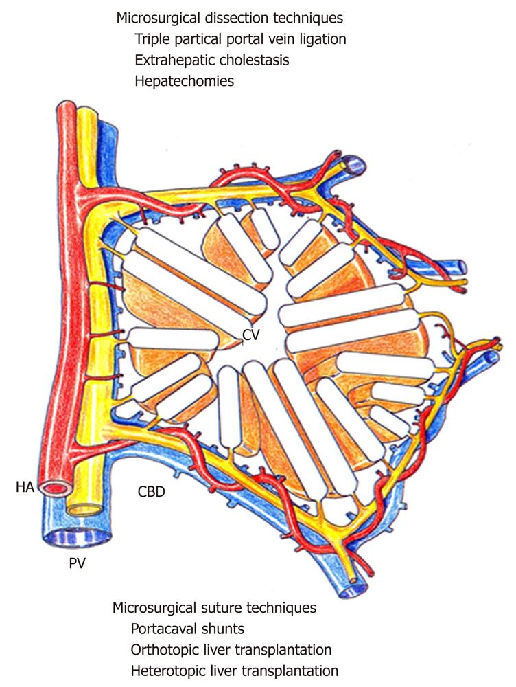
Figure 1 Microsurgical techniques used in liver research.
The liver has been schematically represented as a hepatic functional unit formed by parenchyma (hepatic trabeculae) and stroma (portal space with vascular and biliary structures). The microsurgical experimental models are listed around this hepatic functional unit, grouped according the predominantly surgical technique used. Thus, hilar and parenchymal microdissection and vascular and biliary microsutures are the two fundamental microsurgical techniques used to obtain experimental models of liver diseases. Therefore, both microsurgical techniques can be considered for classifying the experimental models developed. HA: Hepatic artery; CBD: Common bile duct; PV: Portal vein; CV: Centrolobular vein.
- Citation: Aller MA, Arias N, Prieto I, Agudo S, Gilsanz C, Lorente L, Arias JL, Arias J. A half century (1961-2011) of applying microsurgery to experimental liver research. World J Hepatol 2012; 4(7): 199-208
- URL: https://www.wjgnet.com/1948-5182/full/v4/i7/199.htm
- DOI: https://dx.doi.org/10.4254/wjh.v4.i7.199


