Copyright
©2012 Baishideng Publishing Group Co.
World J Hepatol. Nov 27, 2012; 4(11): 314-318
Published online Nov 27, 2012. doi: 10.4254/wjh.v4.i11.314
Published online Nov 27, 2012. doi: 10.4254/wjh.v4.i11.314
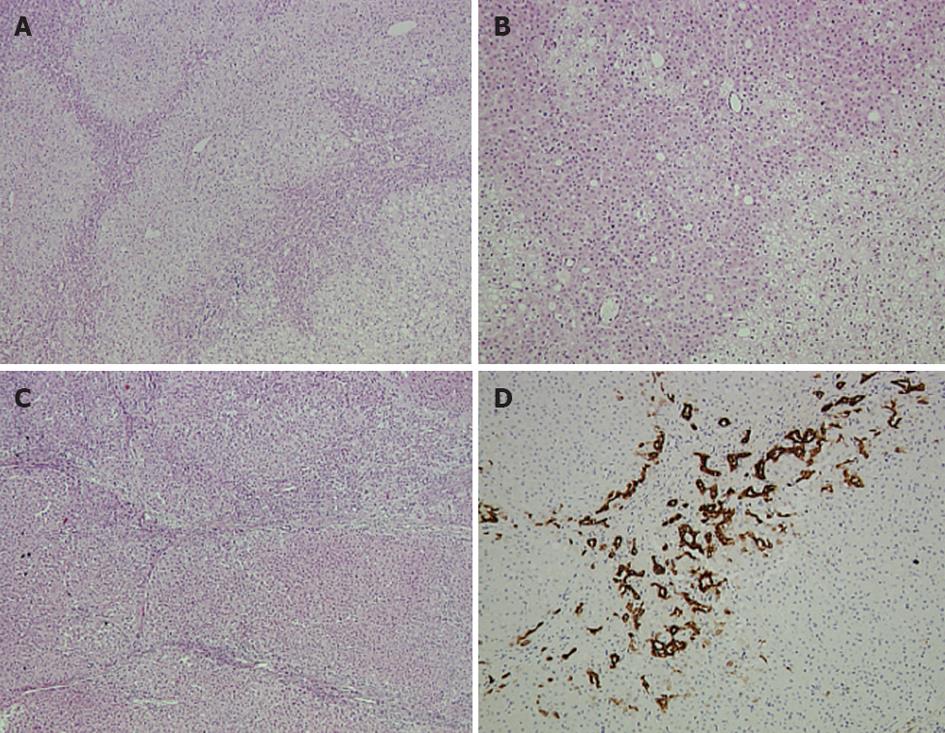
Figure 2 Pathology report of the larger lesion in segments.
A, B: Hematoxylin and eosin (HE) staining of the hepatocellular adenoma showing a vague lobularity created by two hepatocytic populations with zonal arrangement. Rosette-like formations are apparent (A ×5, B ×10); C: HE staining of the focal nodular hyperplasia lesion showing nodularity and thin fibrous septa (×5); D: Ductular reaction depicted by cytokeratin 7 expression (×10).
- Citation: Dimitroulis D, Lainas P, Charalampoudis P, Karatzas T, Delladetsima I, Sakellariou S, Karidis N, Kouraklis G. Co-existence of hepatocellular adenoma and focal nodular hyperplasia in a young female. World J Hepatol 2012; 4(11): 314-318
- URL: https://www.wjgnet.com/1948-5182/full/v4/i11/314.htm
- DOI: https://dx.doi.org/10.4254/wjh.v4.i11.314


