Copyright
©2012 Baishideng Publishing Group Co.
World J Hepatol. Oct 27, 2012; 4(10): 284-287
Published online Oct 27, 2012. doi: 10.4254/wjh.v4.i10.284
Published online Oct 27, 2012. doi: 10.4254/wjh.v4.i10.284
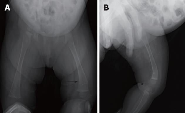
Figure 5 Plain skeletal radiographic features at the 28 d after hepaticojejunostomy in the case 2.
Anteroposterior (A) and lateral (B) plain radiographs showing a displaced fracture (arrows) of the left distal femur.
- Citation: Okada T, Honda S, Miyagi H, Minato M, Taketomi A. Hepatic osteodystrophy complicated with bone fracture in early infants with biliary atresia. World J Hepatol 2012; 4(10): 284-287
- URL: https://www.wjgnet.com/1948-5182/full/v4/i10/284.htm
- DOI: https://dx.doi.org/10.4254/wjh.v4.i10.284


