Copyright
©2012 Baishideng Publishing Group Co.
World J Hepatol. Oct 27, 2012; 4(10): 284-287
Published online Oct 27, 2012. doi: 10.4254/wjh.v4.i10.284
Published online Oct 27, 2012. doi: 10.4254/wjh.v4.i10.284
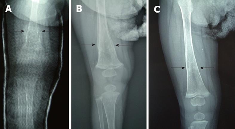
Figure 3 Plain skeletal radiographic features at the 8 d after the application of an immobilizing plaster bandage for the femur fracture in the case 1.
Callus formation (arrows) was seen 8 d after the application of an immobilizing plaster bandage (A) in case 1. The plaster bandage was removed after 20 d (B) and the fracture of the right femur was cured 6 mo post-fracture (C).
- Citation: Okada T, Honda S, Miyagi H, Minato M, Taketomi A. Hepatic osteodystrophy complicated with bone fracture in early infants with biliary atresia. World J Hepatol 2012; 4(10): 284-287
- URL: https://www.wjgnet.com/1948-5182/full/v4/i10/284.htm
- DOI: https://dx.doi.org/10.4254/wjh.v4.i10.284


