Copyright
©2011 Baishideng Publishing Group Co.
World J Hepatol. Sep 27, 2011; 3(9): 256-261
Published online Sep 27, 2011. doi: 10.4254/wjh.v3.i9.256
Published online Sep 27, 2011. doi: 10.4254/wjh.v3.i9.256
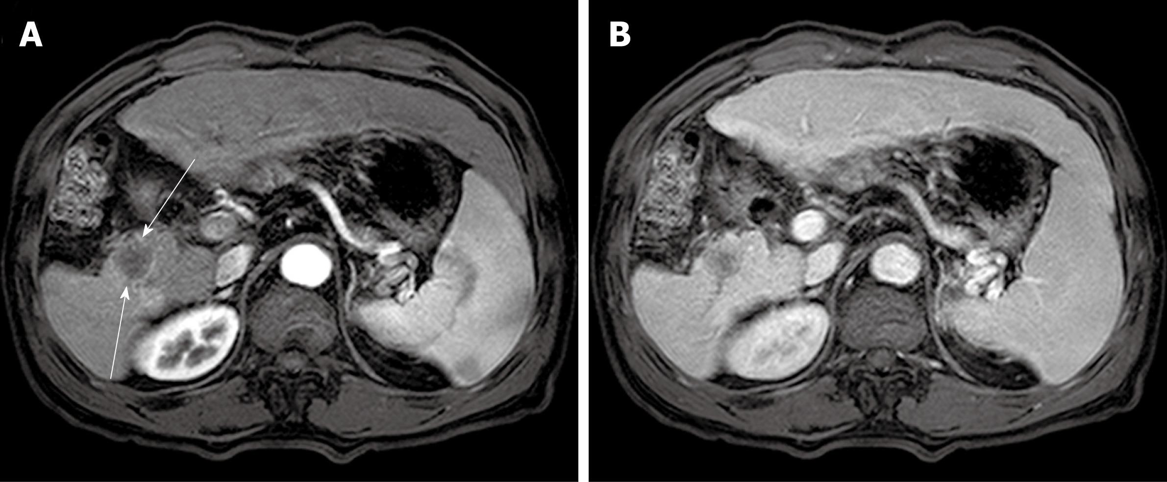
Figure 4 Computed tomography scan at follow-up of twenty-one months.
A: A well demarcated low-attenuated round mass was newly found at the resection margin of right posterior segment (slender arrows). This mass was not significantly enhanced in late arterial phase; B: This mass was also not well enhanced at portal phase, which is similar pattern with the previous malignant fibrous histiocytoma.
- Citation: Hwang HS, Ha ND, Jeong YK, Suh JH, Choi HJ, Kim YM, Cha HJ. Simultaneous occurrence of malignant fibrous histiocytoma and hepatocellular carcinoma in cirrhotic liver: A case report. World J Hepatol 2011; 3(9): 256-261
- URL: https://www.wjgnet.com/1948-5182/full/v3/i9/256.htm
- DOI: https://dx.doi.org/10.4254/wjh.v3.i9.256


