Copyright
©2011 Baishideng Publishing Group Co.
World J Hepatol. Jul 27, 2011; 3(7): 184-197
Published online Jul 27, 2011. doi: 10.4254/wjh.v3.i7.184
Published online Jul 27, 2011. doi: 10.4254/wjh.v3.i7.184
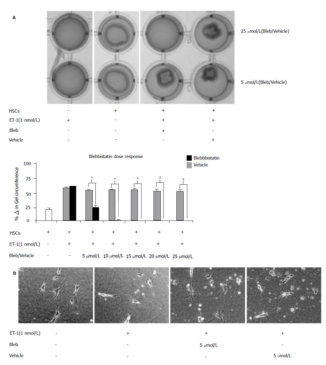
Figure 7 Blebbistatin-inhibited endothelin-1-induced hepatic stellate cell contraction.
Culture-activated hepatic stellate cells(HSCs) (Day 4) were serum-starved 24 h prior to seeding onto collagen lattices (3 × 10⁵ cells/well). Cells were pretreated with inactive (Vehicle) or active (Blebbistatin) myosin II inhibitor in increasing doses (0-25 μmol/L; 5) and subsequently treated with blebbistatin-inhibited endothelin-1 (ET-1) (1 nmol/L) after 30 min. (aP < 0.05 as compared to scramble). A: Representative light micrographs of collagen lattices: with or without seeded HSCs; with or without chemical treatments (top panel). Twenty-four hours following chemical treatment, hepatic stellate cell contraction was quantified using PTI ImageMaster software and reported as percentage change in gel circumference (bottom panel). B: Representative light micrographs of collagen-seeded HSCs with or without chemical treatments.
- Citation: Moore CC, Lakner AM, Yengo CM, Schrum LW. Nonmuscle myosin II regulates migration but not contraction in rat hepatic stellate cells. World J Hepatol 2011; 3(7): 184-197
- URL: https://www.wjgnet.com/1948-5182/full/v3/i7/184.htm
- DOI: https://dx.doi.org/10.4254/wjh.v3.i7.184


