Copyright
©2010 Baishideng.
World J Hepatol. Apr 27, 2010; 2(4): 151-161
Published online Apr 27, 2010. doi: 10.4254/wjh.v2.i4.151
Published online Apr 27, 2010. doi: 10.4254/wjh.v2.i4.151
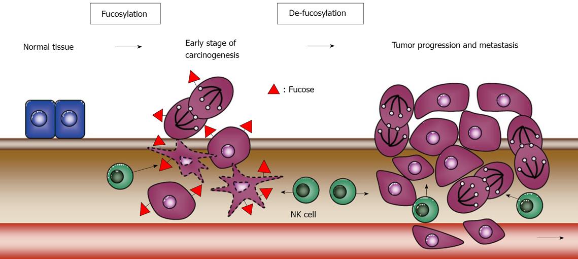
Figure 4 Schematic model of the biological function of fucosylation and de-fucosylation in modulating immune surveillance during colon carcinogenesis[109].
The level of fucosylation is not high in normal colon tissues, but is increased at an early stage in colon cancer. The cancer cells represented by the dotted line are apoptotic ones, which are attacked by NK cells. In certain types of advanced cancer, de-fucosylation through genetic mutation leads to escape from NK cell-mediated tumor surveillance and the acquisition of more malignant characteristics. This figure is modified from the data in reference 109.
- Citation: Moriwaki K, Miyoshi E. Fucosylation and gastrointestinal cancer. World J Hepatol 2010; 2(4): 151-161
- URL: https://www.wjgnet.com/1948-5182/full/v2/i4/151.htm
- DOI: https://dx.doi.org/10.4254/wjh.v2.i4.151


