Copyright
©2010 Baishideng.
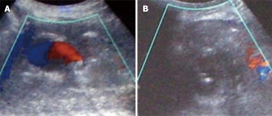
Figure 2 Ultrasound guided thrombin injection.
A: Color doppler ultrasound. A small cavity persists with flow in the pseudoaneurysm; B: Needle inside the pseudoaneurysm immediately after the injection of thrombin showing the absence of flow (thrombosis).
- Citation: Francisco LE, Asunción LC, Antonio CA, Ricardo RC, Manuel RP, Caridad MH. Post-traumatic hepatic artery pseudoaneurysm treated with endovascular embolization and thrombin injection. World J Hepatol 2010; 2(2): 87-90
- URL: https://www.wjgnet.com/1948-5182/full/v2/i2/87.htm
- DOI: https://dx.doi.org/10.4254/wjh.v2.i2.87


