Copyright
©2010 Baishideng Publishing Group Co.
World J Hepatol. Nov 27, 2010; 2(11): 410-415
Published online Nov 27, 2010. doi: 10.4254/wjh.v2.i11.410
Published online Nov 27, 2010. doi: 10.4254/wjh.v2.i11.410
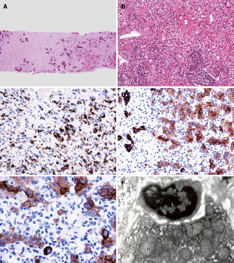
Figure 1 Liver biopsy showed extensive patchy areas of multilobular necrosis with only bile ducts remaining, extensive ductal metaplasia, severe lymphocytic and macrophages infiltration of portal tracts and lobular parenchyma and patchy plasma cell infiltrates.
Histological changes were consistent with acute troxis necrosis and fulminant hepatitis. A: Liver lobules showing massive necrosis with only bile ducts remaining (hematoxiline and eosin stain × 52); B: Lymphocytic infiltration of portal tract and lobular parenchyma (hematoxiline and eosin stain × 130); C: Liver lobular necrosis with macrophages cleaning the debris (CD68 stain × 130); D: Ductal metaplasia. Lymphocytic infiltration in the sinusoids (CAM5.2 stain × 260); E: High power, lymphocytes destroying hepatocytes (CAM5.2 stain × 520); F: Lymphocyte “eating” hepatocytes in a liver parenchyma (troxis necrosis), arrow showing immunological synapses (Electron microscopy × 15000).
- Citation: Chen GC, Ramanathan VS, Law D, Funchain P, Chen GC, French S, Shlopov B, Eysselein V, Chung D, Reicher S, Pham BV. Acute liver injury induced by weight-loss herbal supplements. World J Hepatol 2010; 2(11): 410-415
- URL: https://www.wjgnet.com/1948-5182/full/v2/i11/410.htm
- DOI: https://dx.doi.org/10.4254/wjh.v2.i11.410


