Copyright
©2010 Baishideng Publishing Group Co.
World J Hepatol. Oct 27, 2010; 2(10): 384-386
Published online Oct 27, 2010. doi: 10.4254/wjh.v2.i10.384
Published online Oct 27, 2010. doi: 10.4254/wjh.v2.i10.384
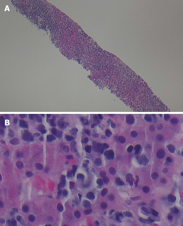
Figure 2 Views of liver biopsy demonstrating atypical mononucleate cells with very high nucleus/cytoplasmic ratios; hyperchromatic with irregularly distributed chromatin; and, irregular nuclear membranes.
A: Low power view; B: High power view. Some nucleoli are infiltrating throughout the sinusoidal spaces. These are features of malignancy. The benign hepatocytes show eosinophilic granular cytoplasm and centrally placed round regular nucleus.
- Citation: Davis ML, Hashemi N. Acute liver failure as a rare initial manifestation of peripheral T-cell lymphoma. World J Hepatol 2010; 2(10): 384-386
- URL: https://www.wjgnet.com/1948-5182/full/v2/i10/384.htm
- DOI: https://dx.doi.org/10.4254/wjh.v2.i10.384


