
















©2010 Baishideng Publishing Group Co.
World J Hepatol. Oct 27, 2010; 2(10): 384-386
Published online Oct 27, 2010. doi: 10.4254/wjh.v2.i10.384
Published online Oct 27, 2010. doi: 10.4254/wjh.v2.i10.384
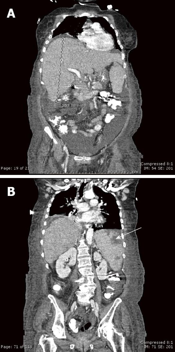
Figure 1 Contrast-enhanced computed tomography coronal reconstructions.
A: An enlarged, heterogenous liver, 20 cm in the craniocaudad dimension; B: There are also multiple, subcapsular peripheral defects that lack enhancement in an enlarged spleen (arrow) representing splenic infarcts. Other computed tomography findings (not depicted) included mediastinal, right hilar, and chest wall lymphadenopathy as well as moderate ascites.
- Citation: Davis ML, Hashemi N. Acute liver failure as a rare initial manifestation of peripheral T-cell lymphoma. World J Hepatol 2010; 2(10): 384-386
- URL: https://www.wjgnet.com/1948-5182/full/v2/i10/384.htm
- DOI: https://dx.doi.org/10.4254/wjh.v2.i10.384