
















Copyright: ©Author(s) 2026.
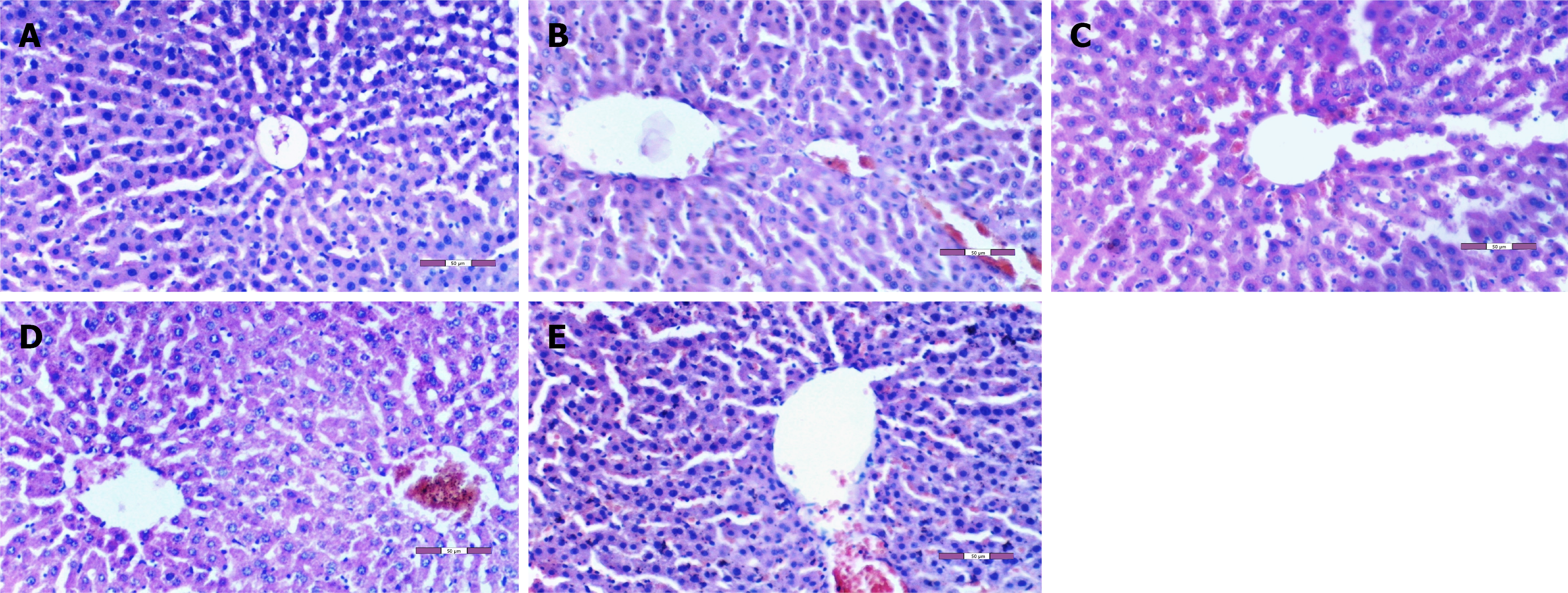
Figure 2 Representative hematoxylin and eosin-stained liver section.
A: Control group (group C): Normal hepatic architecture is preserved, with well-organized hepatocyte cords, intact sinusoidal spaces, and normal nuclear morphology. No evidence of hepatocellular degeneration, inflammatory cell infiltration, or ischemia-reperfusion injury (IRI)-related tissue injury is observed (× 200); B: IRI group: Marked hepatic injury is evident, characterized by hepatocellular degeneration, prominent sinusoidal dilatation, pyknotic nuclei, and parenchymal mononuclear inflammatory cell infiltration, consistent with severe IRI-induced tissue damage (× 200); C: Desflurane-treated IRI group: Histopathological injury is less pronounced than in the untreated IRI group, with reduced hepatocellular degeneration and sinusoidal dilatation accompanied by mild inflammatory cell infiltration, indicating partial preservation of hepatic tissue architecture (× 200); D: Fullerenol C60-treated IRI group: Liver architecture is largely preserved, with only mild hepatocellular degeneration, limited sinusoidal dilatation, and minimal inflammatory cell infiltration, demonstrating attenuation of IRI-induced histopathological injury (× 200); E: Combined fullerenol C60 and desflurane-treated IRI group: Mild to moderate histopathological alterations, including limited hepatocellular degeneration, sinusoidal dilatation, and scattered inflammatory cell infiltration, are observed. Overall tissue injury is markedly reduced compared with the untreated IRI group and is comparable to that observed in the fullerenol-treated group (× 200).
- Citation: Kasapbasi E, Ozdemir C, Yavuz A, Sezen SC, Kurtipek O, Kavutçu M, Arslan M. Effects of fullerenol C60 on hepatic ischemia-reperfusion injury in desflurane-anesthetized rats. World J Hepatol 2026; 18(9): 122845
- URL: https://www.wjgnet.com/1948-5182/full/v18/i9/122845.htm
- DOI: https://dx.doi.org/10.4254/wjh.122845