
















Copyright: ©Author(s) 2026.
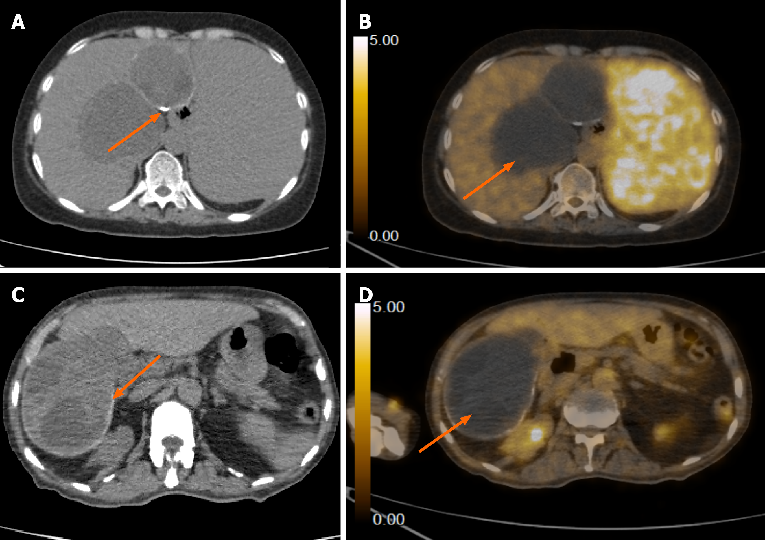
Figure 14 Positron emission tomography/computed tomography images of hydatid cysts in two patients.
A: Plain computed tomography (CT) axial image showing a cystic lesion with wall calcification (arrow); B: Fused positron emission tomography/CT axial image shows no evidence of fluorodeoxyglucose activity within the lesion (arrow); C: Plain CT axial image shows another hydatid cyst in segment VI of the liver with peripheral calcification (arrow); D: Fused positron emission tomography/CT axial image shows no corresponding fluorodeoxyglucose activity (arrow).
- Citation: Khandwala K, Sajjad M, Hussain Z, Zeeshan M, Hilal K. Hepatic hydatid cyst disease in low- and middle-income countries: Spectrum of imaging findings and complications. World J Hepatol 2026; 18(8): 123946
- URL: https://www.wjgnet.com/1948-5182/full/v18/i8/123946.htm
- DOI: https://dx.doi.org/10.4254/wjh.123946