
















Copyright: ©Author(s) 2026.
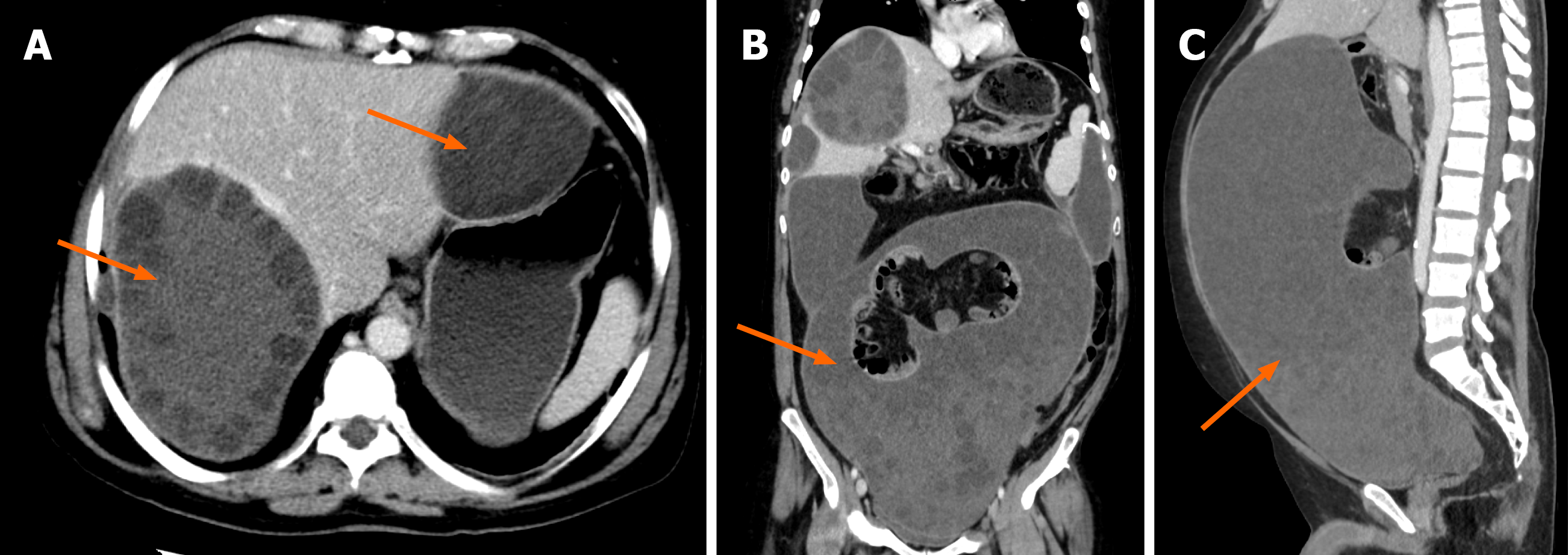
Figure 11 Hydatid cyst disease with intraperitoneal dissemination.
A-C: Axial, coronal and sagittal computed tomography images showing at least two hydatid cysts with daughter cysts with a typical rosette appearance in the left and right lobes of the liver (arrows in A). Coronal and sagittal sections show diffuse dissemination of hydatid disease into the peritoneum (B and C).
- Citation: Khandwala K, Sajjad M, Hussain Z, Zeeshan M, Hilal K. Hepatic hydatid cyst disease in low- and middle-income countries: Spectrum of imaging findings and complications. World J Hepatol 2026; 18(8): 123946
- URL: https://www.wjgnet.com/1948-5182/full/v18/i8/123946.htm
- DOI: https://dx.doi.org/10.4254/wjh.123946