
















Copyright: ©Author(s) 2026.
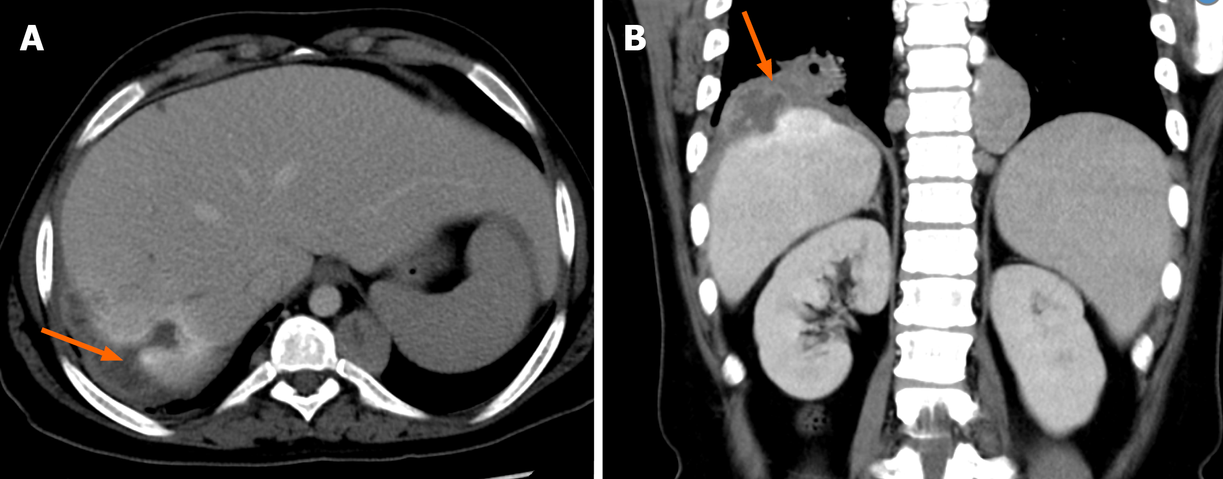
Figure 10 Hydatid cyst disease with rupture and transdiaphragmatic spread.
A and B: Axial computed tomography (A) and coronal computed tomography (B) images showing subcapsular rupture of hydatid disease in the right lobe of the liver involving segment VII (arrow in A) with transdiaphragmatic extension into the ipsilateral right thoracic cavity (arrow in B).
- Citation: Khandwala K, Sajjad M, Hussain Z, Zeeshan M, Hilal K. Hepatic hydatid cyst disease in low- and middle-income countries: Spectrum of imaging findings and complications. World J Hepatol 2026; 18(8): 123946
- URL: https://www.wjgnet.com/1948-5182/full/v18/i8/123946.htm
- DOI: https://dx.doi.org/10.4254/wjh.123946