
















Copyright: ©Author(s) 2026.
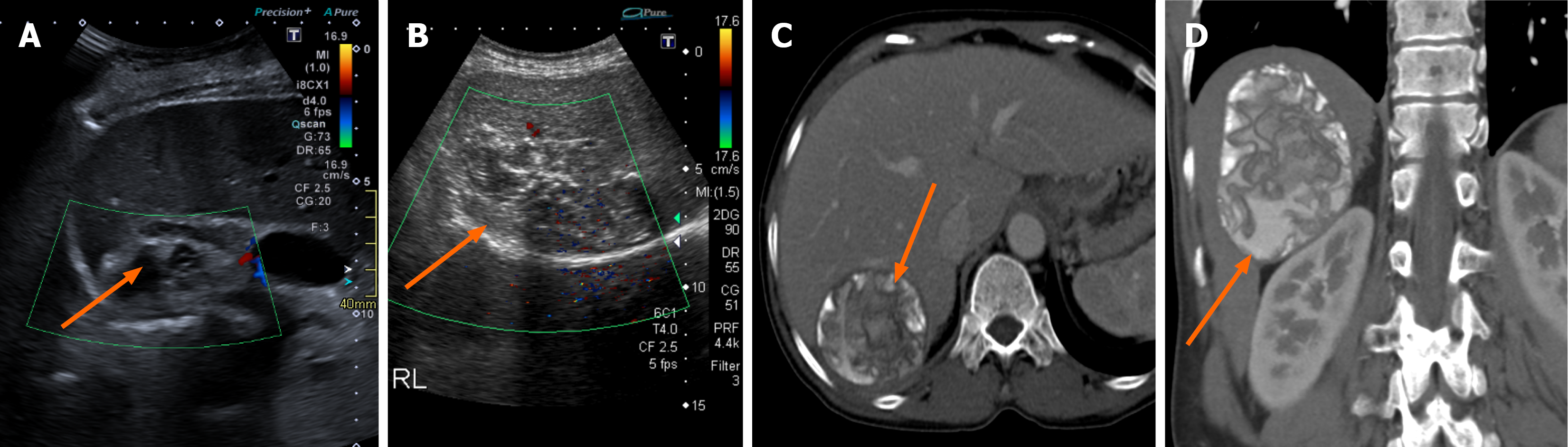
Figure 5 Cystic echinococcosis 4 hydatid cysts.
A and B: Ultrasound images from two different patients showing a cystic echinococcosis 4 degenerative hydatid cyst in the right lobe of the liver (arrows) with a heterogeneous appearance on ultrasound with mixed echoes and no definite daughter vesicles; C and D: Axial (C) and coronal (D) images reveal the characteristic cystic echinococcosis 4 hydatid cyst with crumpled membranes and calcifications (arrows).
- Citation: Khandwala K, Sajjad M, Hussain Z, Zeeshan M, Hilal K. Hepatic hydatid cyst disease in low- and middle-income countries: Spectrum of imaging findings and complications. World J Hepatol 2026; 18(8): 123946
- URL: https://www.wjgnet.com/1948-5182/full/v18/i8/123946.htm
- DOI: https://dx.doi.org/10.4254/wjh.123946