
















Copyright: ©Author(s) 2026.
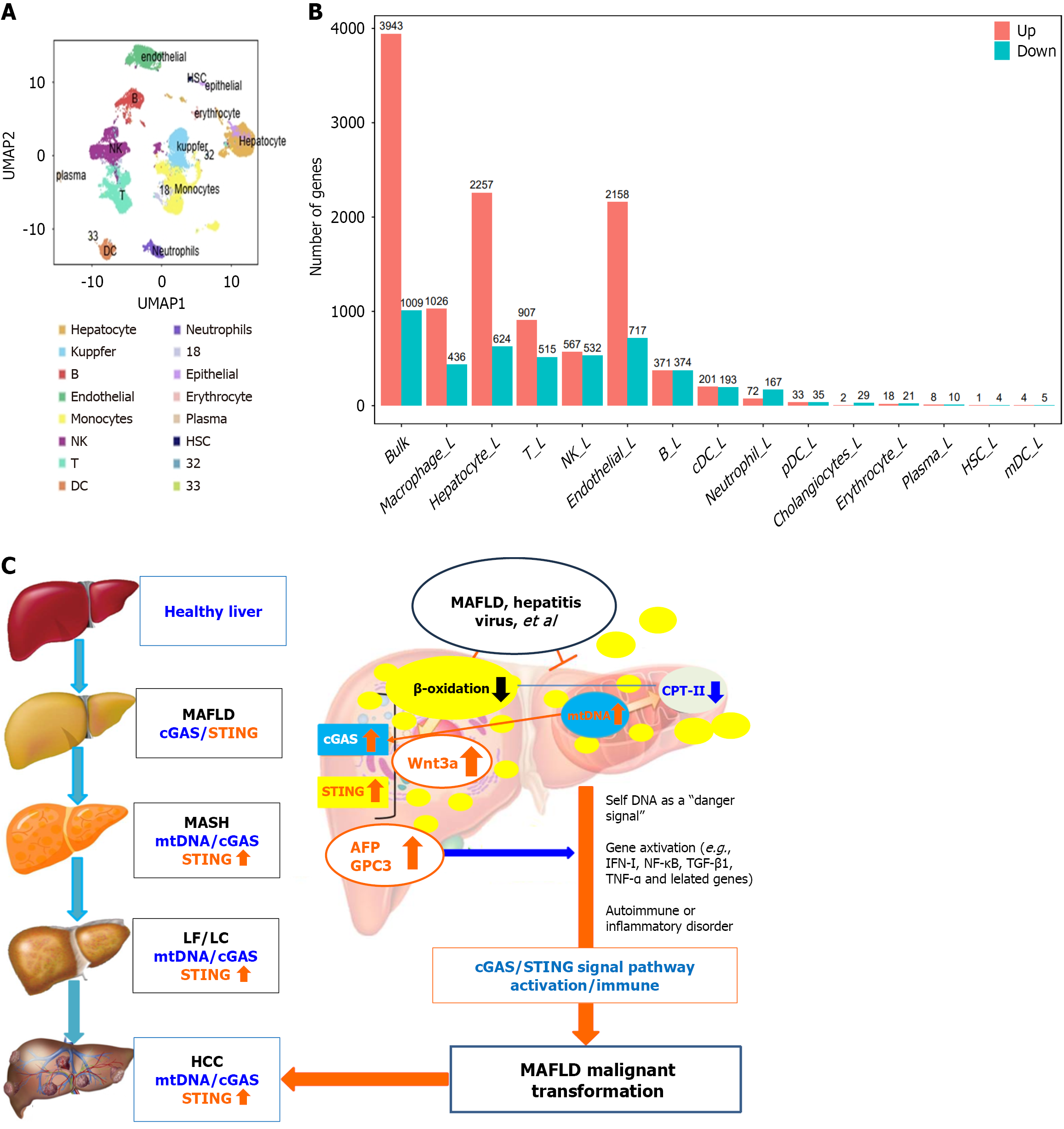
Figure 6 Alterations in immune cells and a possible mechanism of cyclic guanosine monophosphate-adenosine monophosphate synthase-stimulator of interferon genes activation.
A: Clustering of liver immune cells. The cell types identified were hepatocytes, B cells, endothelial cells, natural killer cells, T cells, monocytes, dendritic cells, neutrophils, epithelial cells, red blood cells, plasma cells, hepatocytes, and dendritic cells. Unidentified cells (8, 32, 33); B: Gene transcription of immune cells in metabolic dysfunction-associated fatty liver disease; C: A possible mechanism by which metabolic dysfunction-associated fatty liver disease malignancy is promoted. NK: Natural killer; DC: Dendritic cell; HSC: Hepatic stellate cells; UMAP: Uniform manifold approximation and projection; cGAS: Cyclic guanosine monophosphate-adenosine monophosphate synthase; STING Stimulator of interferon genes; mtDNA: Mitochondrial DNA; LF: Liver fibrosis; AFP: Alpha-fetoprotein; GPC3: Glypican-3; CPT-II: Carnitine palmitoyl transferase II; INF-I: Interferon-I; NF-κB: Nuclear factor kappa-B; TGF-β1: Transforming growth factor β1; TNF-α: Tumor necrosis factor α; MAFLD: Metabolic dysfunction-associated fatty liver disease; LC: Liver cirrhosis.
- Citation: Zhou MY, Fang RF, Tang H, Xia XX, Xie Q, Yao DF, Sai WL, Yao M. Activated cGAS-STING signaling promotes malignancy in metabolic dysfunction-associated fatty liver disease via mitochondrial DNA and immune cell dysfunction. World J Hepatol 2026; 18(7): 121423
- URL: https://www.wjgnet.com/1948-5182/full/v18/i7/121423.htm
- DOI: https://dx.doi.org/10.4254/wjh.121423