
















Copyright: ©Author(s) 2026.
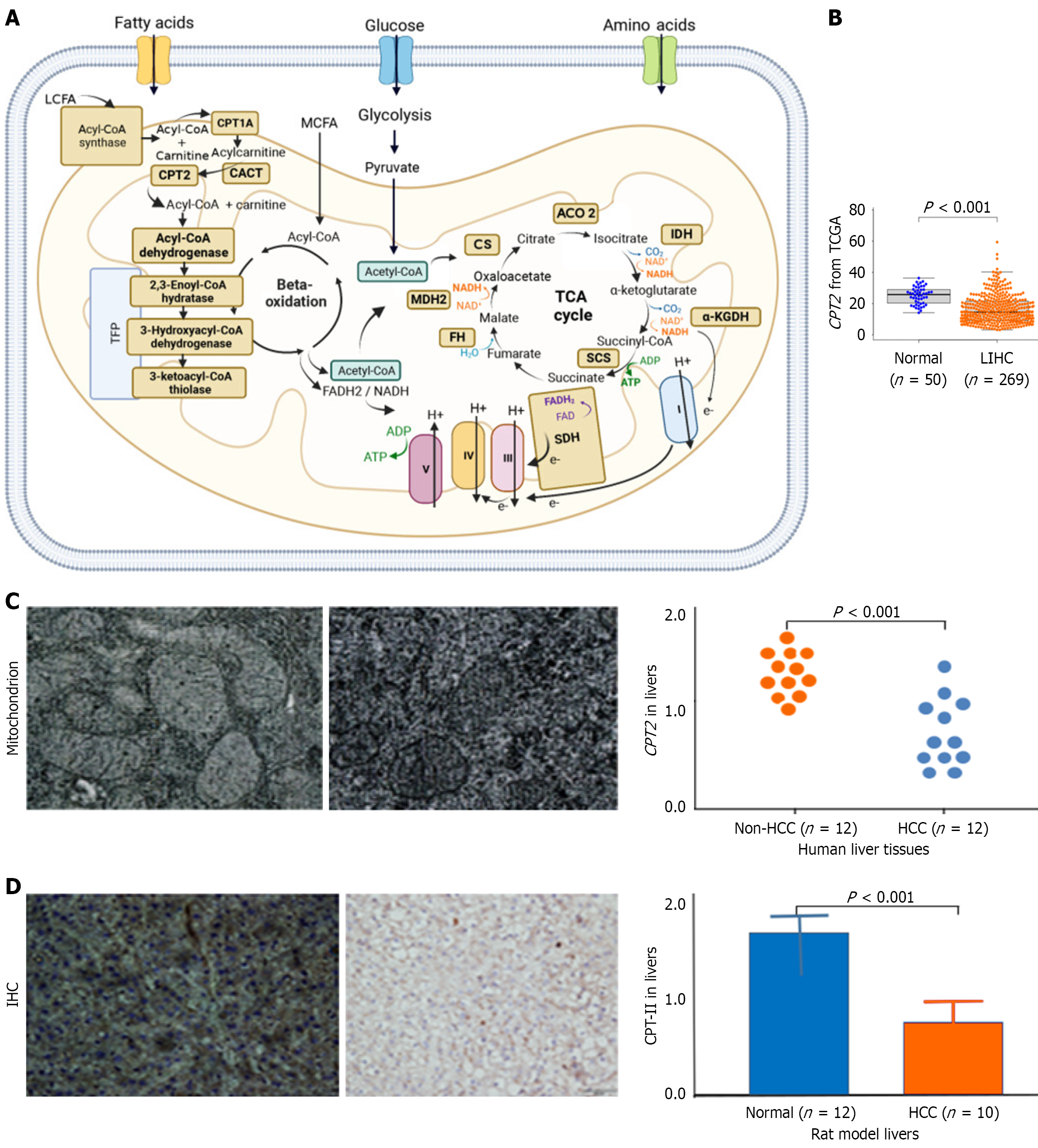
Figure 2 Mitochondrial damage and CPT2/CPT-II during lipid accumulation.
A: CPT2 located on the mitochondrial membrane; B: Comparison of liver CPT2 expressions between healthy liver (n = 50) and hepatocellular carcinoma (HCC) tissues (n = 269, liver hepatocellular carcinoma) from The Cancer Genome Atlas database; C: Damaged mitochondria and CPT2 expressions in human liver tissues: (1) Upper left: Mitochondria in non-HCC (n = 12); and (2) Upper right: Damaged mitochondria in HCC tissues (n = 12); D: CPT-II expressions between healthy liver (n = 12) and HCC (n = 10) tissues from the rat models. cP < 0.001 vs the NC or noncancerous group. LCFA: Long-chain fatty acid; CPT2: Carnitine palmitoyl transferase II gene; CPT1A: Carnitine palmitoyl transferase 1a; CACT: Carnitine acylcarnitine translocase; Acyl-CoA: Acyl coenzyme A; ACO2: Aconitase 2; IDH: Isocitrate dehydrogenase; NAD: Nicotinamide adenine dinucleotide; NADH: Nicotinamide adenine dinucleotide-1; α-KGDH: Α-Ketoglutarate dehydrogenase; MDH2: Malate dehydrogenase 2; TCA: Tricarboxylic acid cycle; FADH1: Nicotinamide adenine dinucleotide phosphate1; SDH: Saccharopine dehydrogenase; ATP: Adenosine triphosphate; ADP: Adenosine diphosphate; TCGA: The Cancer Genome Atlas; CPT-II: Carnitine palmitoyl transferase II; IHC: Immunohistochemistry; HCC: Hepatocellular carcinoma; LIHC: Liver hepatocellular carcinoma.
- Citation: Zhou MY, Fang RF, Tang H, Xia XX, Xie Q, Yao DF, Sai WL, Yao M. Activated cGAS-STING signaling promotes malignancy in metabolic dysfunction-associated fatty liver disease via mitochondrial DNA and immune cell dysfunction. World J Hepatol 2026; 18(7): 121423
- URL: https://www.wjgnet.com/1948-5182/full/v18/i7/121423.htm
- DOI: https://dx.doi.org/10.4254/wjh.121423