
















Copyright: ©Author(s) 2026.
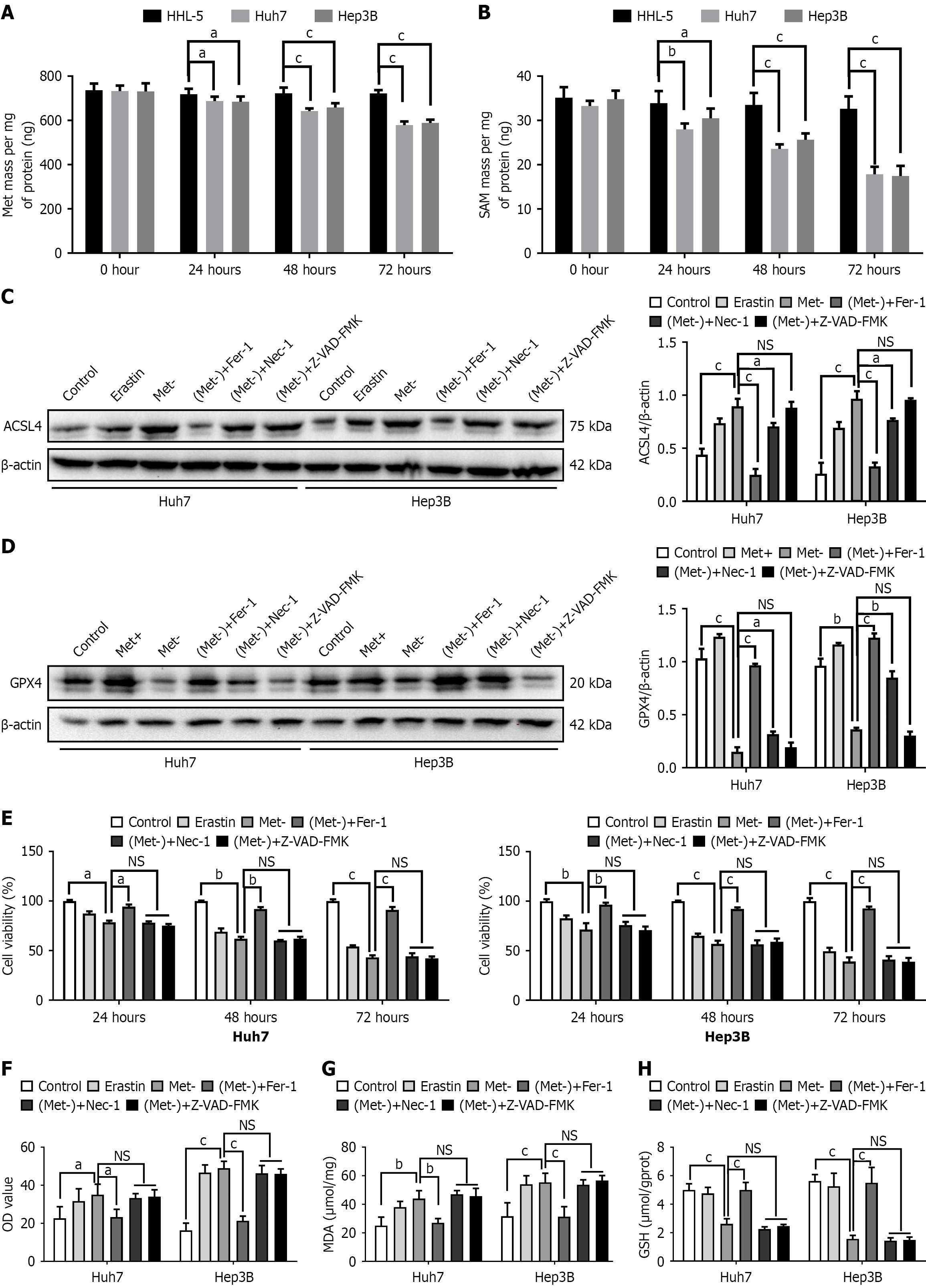
Figure 1 Methionine deficiency promotes hepatocellular carcinoma cell ferroptosis.
A and B: Intracellular methionine and S-adenosylmethionine levels were determined using ELISA assay; C and D: ACSL4 and GPX4 levels were examined by western blot; E: Cell proliferation activity was assessed using the CCK-8 assay; F: Reactive oxygen species levels were measured using the fluorescent probe DCFH-DA; G: Intracellular lipid peroxidation was evaluated using the malondialdehyde assay kit; H: Intracellular glutathione levels were determined using the glutathione colorimetric assay. Erastin (a ferroptosis inducer, 10 μm, 24 hours), ferrostatin-1 (Fer-1, a potent and selective inhibitor of ferroptosis, 2 μm, 24 hours), necrostatin-1 (Nec-1, a potent necroptosis inhibitor that crosses the blood-brain barrier, 60 μm, 24 hours), and Z-VAD-FMK (a pan-caspase inhibitor, 50 μm, 24 hours) were used. Results are presented as the mean ± SD, n = 3. aP < 0.05; bP < 0.01; cP < 0.001. Met: Methionine; SAM: S-adenosylmethionine; Met-: Methionine-deficient; Met+: Methionine-supplemented (100 μm); GSH: Glutathione; MDA: Malondialdehyde; NS: Not significant.
- Citation: Yin DD, Jiang WY, Huang YT, Zhou SP, Zhang YC. Methionine depletion inhibits hepatocellular carcinoma by inducing ferroptosis and suppressing epithelial-mesenchymal transition through the ARNT2/UBE2Z pathway. World J Hepatol 2026; 18(6): 120427
- URL: https://www.wjgnet.com/1948-5182/full/v18/i6/120427.htm
- DOI: https://dx.doi.org/10.4254/wjh.120427