
















Copyright: ©Author(s) 2026.
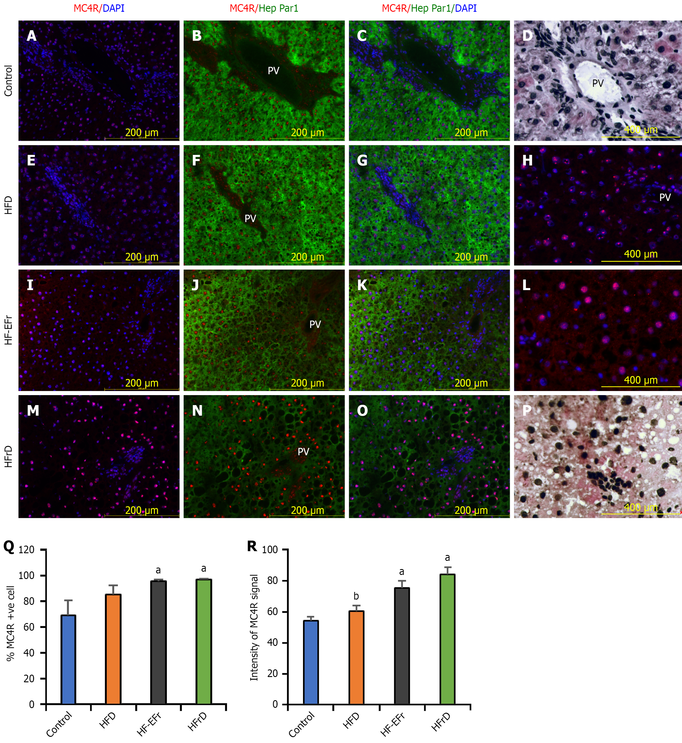
Figure 5 Fructose-enriched diets are associated with prominent nuclear MC4R immunoreactivity in hepatocytes as demonstrated by dual immunofluorescence staining and immunohistochemistry.
A-C: Row represents the control group showing some small, and mainly nuclear positive MC4R red dots [immunofluorescence (IF)]; D: Immunohistochemistry (IHC) at week 8 shows faint cytoplasmic MC4R (brown) in some hepatocytes of formalin-fixed, paraffin-embedded following antigen retrieval. Nuclei are counterstained with hematoxylin (× 400, scale = 50 μm); E-G: High-fat diet (HFD) group micrographs show MC4R (red) and DAPI-stained nuclei (blue); H: Higher magnification (× 400) micrograph at week 8 highlights nuclear MC4R dots; I-K: Represent hepatic micrographs of high-fat plus ethanol and fructose group; MC4R (red) and DAPI (blue) reveal rounded nuclear MC4R positivity; L: Higher magnification (× 400) confirms nuclear localization at week 8; M-O: Micrographs of Lieber-DeCarli high-fructose diet group display the strongest MC4R nuclear positivity and accumulation (IF); P: IHC shows MC4R in both nuclear and cytoplasmic compartments, with numerous clear vacuoles due to lipid droplet loss (steatosis), nuclei were counterstained by hematoxylin (× 400); Q: Percentage of MC4R-positive nuclei at week 4; R: Mean fluorescence intensity (MFI) of nuclear-associated MC4R at week 4. aP < 0.05 vs control, bP < 0.05 vs HFD (one-way ANOVA with Tukey post-hoc test). n = 5 rats per group; about 15 images analyzed per group. Nuclear MC4R immunoreactivity is described at the level of protein localization by IF and IHC; no functional inference regarding nuclear MC4R signaling is made here. HFD: High-fat diet; HF-EFr: High-fat plus ethanol and fructose; HFrD: High-fructose diet; PV: Portal vein.
- Citation: Alwahsh SM, Xu M, Nabulsi M, Mihm S, Alzahrani FA, Ramadori GP. Differential impact of high-fructose and ethanol diets on early steatohepatitis and hepatic melanocortin-4 receptor responses in rats. World J Hepatol 2026; 18(6): 119792
- URL: https://www.wjgnet.com/1948-5182/full/v18/i6/119792.htm
- DOI: https://dx.doi.org/10.4254/wjh.119792