
















Copyright: ©Author(s) 2026.
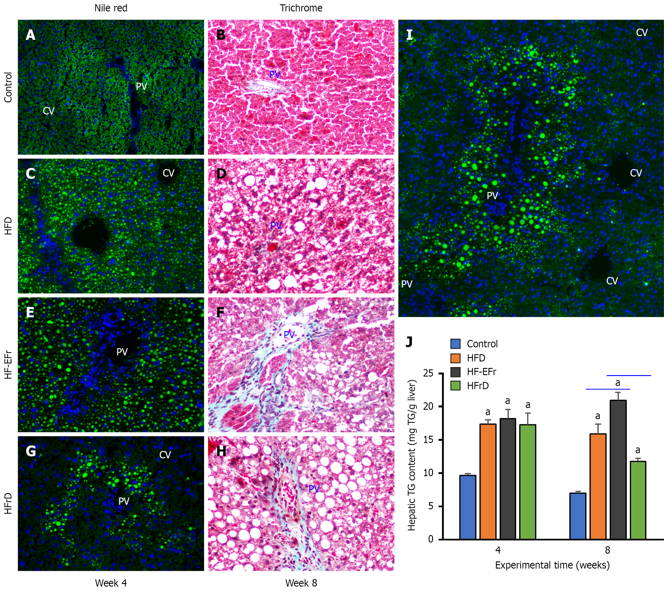
Figure 2 Hepatic steatosis and progressive fibrosis characterization by Nile red (left panel), Masson’s trichrome (right panel) at weeks 4 and 8, respectively, and quantification of hepatic triglyceride content.
A and B: Control rats micrographs show preserved hepatic architecture without lipid accumulation or fibrosis; C and D: High-fat diet group micrographs display diffuse micro- and medio-vesicular steatosis with minimal collagen deposition; E and F: High-fat plus ethanol and fructose (HF-EFr) group micrographs exhibit abundant lipid droplets of variable sizes, with increased portal and perisinusoidal collagen deposition extending into adjacent lobular regions, consistent with stage 2 fibrosis; G and H: Lieber-DeCarli high-fructose diet (HFrD) group micrographs demonstrate marked macro-vesicular steatosis, mainly in the periportal (portal vein) region accompanied by stage 2 fibrosis; Micrographs were acquired at × 20 magnification; I: × 5 magnification of Nile red stain of HFrD rats fed for 2 weeks; J: Biochemical quantification of hepatic triglyceride (TG) content (mg TG/g liver homogenate) at weeks 4 and 8. aP < 0.05 vs control. TG content in the liver HF-EFr at week 8 is significant over all groups. HFD: High-fat diet; HF-EFr: High-fat plus ethanol and fructose; HFrD: High-fructose diet.
- Citation: Alwahsh SM, Xu M, Nabulsi M, Mihm S, Alzahrani FA, Ramadori GP. Differential impact of high-fructose and ethanol diets on early steatohepatitis and hepatic melanocortin-4 receptor responses in rats. World J Hepatol 2026; 18(6): 119792
- URL: https://www.wjgnet.com/1948-5182/full/v18/i6/119792.htm
- DOI: https://dx.doi.org/10.4254/wjh.119792