
















Copyright: ©Author(s) 2026.
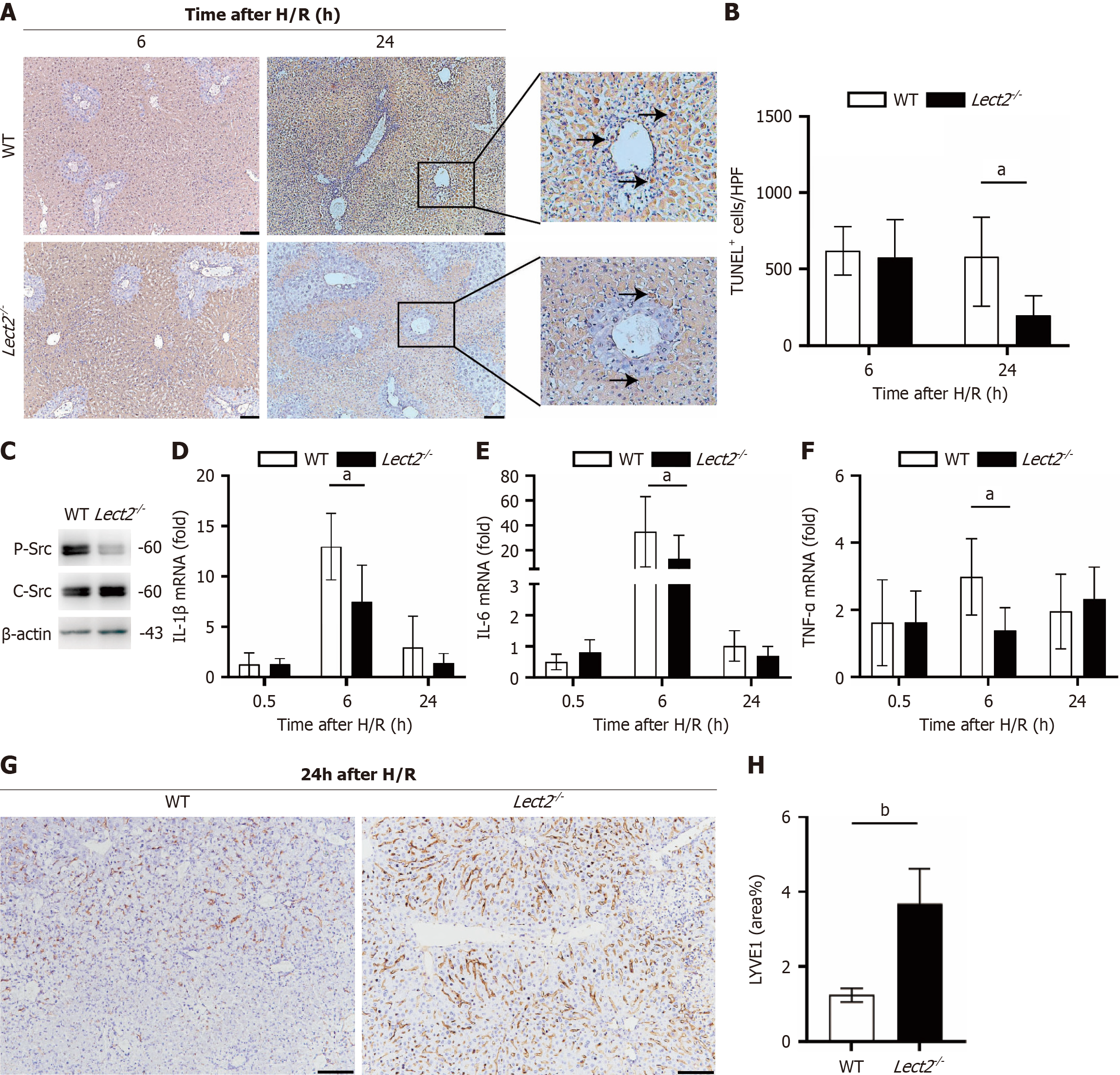
Figure 4 Lect2-/- mice exhibit reduced hepatic ischemia-reperfusion injury and sinusoidal endothelial cell damage.
Livers from Lect2-/- mice and their littermate controls were clamped with the hepatic pedicle for 1 hour followed by reperfusion. Serum and liver tissue samples were collected at the indicated time points. A: TUNEL staining of livers showed apoptosis. Bar, 100 mm; B: Quantification of apoptosis. Student’s t-test; C: Liver tissue lysates from Lect2-/- mice and littermate wild type controls were collected 6 hours after hepatic ischemia-reperfusion injury (1 hour clamping/6 hours reperfusion) and immunoblotted with indicated antibodies; D-F: MRNA levels of interleukin-1β, interleukin-6, and tumor necrosis factor-α in liver tissues. Student’s t-test; G: Lymphatic vessel endothelial hyaluronic acid receptor 1 staining of livers showed apoptosis. Bar, 100 mm; H: Quantification of cell lymphatic vessel endothelial hyaluronic acid receptor 1 staining. Data are representative of three independent experiments. Student’s t-test. n = 6/group. All data are presented as the mean ± SEM. aP < 0.05, bP < 0.01. Lect2: Leukocyte-derived chemotaxin-2; WT: Wild type; H/R: Hypoxia/reoxygenation; IL: Interleukin; TNF-α: Tumor necrosis factor-α; LYVE1: Lymphatic vessel endothelial hyaluronic acid receptor 1.
- Citation: Yang Y, Wu SL, Huang ZH, Dong MQ, Liu ZM, Xu M, Gao Y, Zhou WJ, Lin Y. LECT2/Tie1/Src signaling regulates the oxidative stress response in endothelial cells and liver ischemia-reperfusion injury. World J Hepatol 2026; 18(6): 119561
- URL: https://www.wjgnet.com/1948-5182/full/v18/i6/119561.htm
- DOI: https://dx.doi.org/10.4254/wjh.119561