
















Copyright: ©Author(s) 2026.
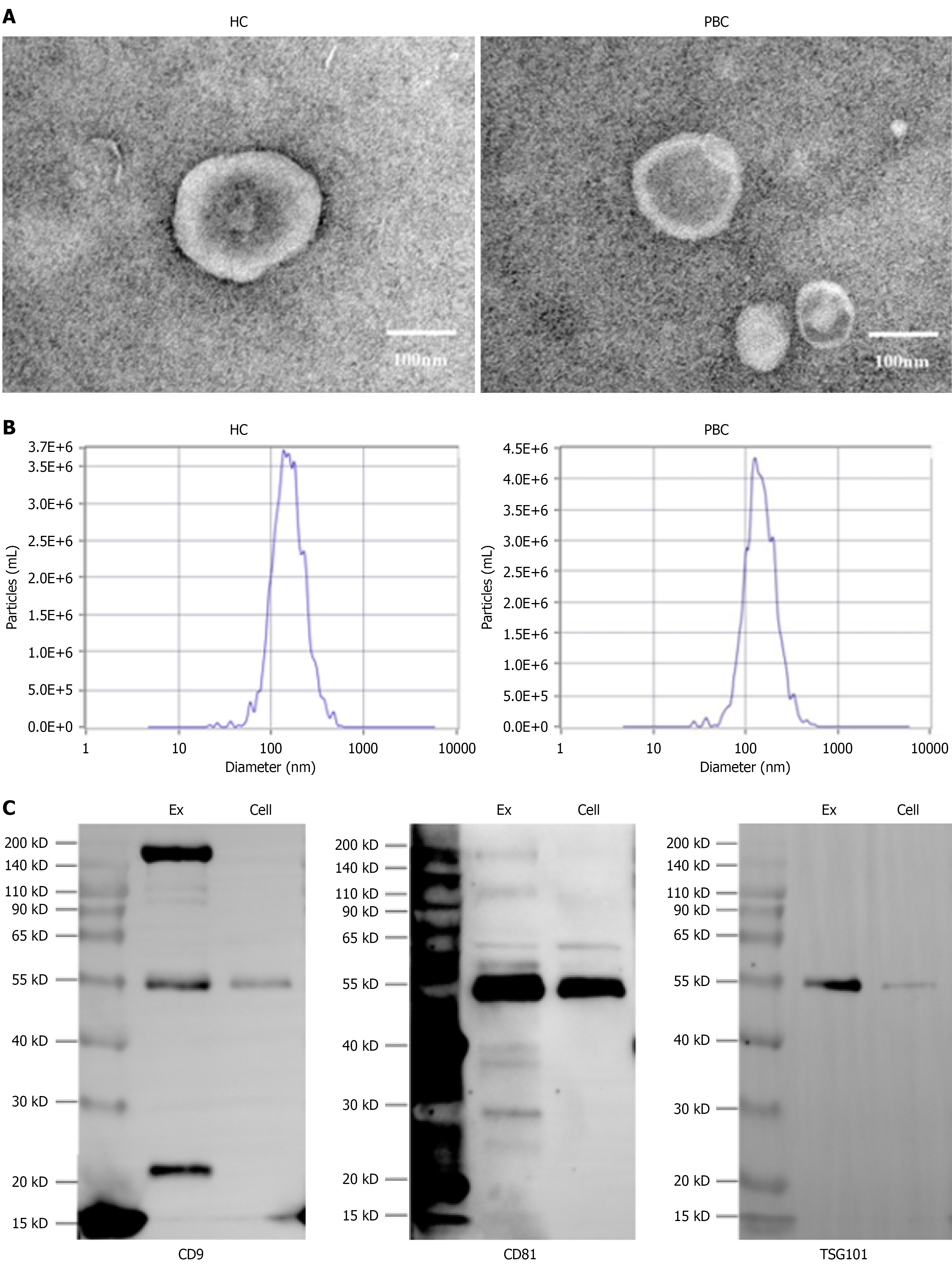
Figure 2 Detection of isolated exosomes.
A: Transmission electron microscopy analysis of exosome preparations from primary biliary cholangitis (PBC) and healthy controls (HCs), representative transmission electron microscopy images of exosomes isolated from plasma of patients with PBC and HCs, showing a discernibly higher particle concentration in PBC samples; B: Nanoparticle tracking analysis indicate that exosomes from both PBC and HCs are similar in having an average diameter of 80-160 nm; C: Immunoblot showed extracted exosomes with significantly higher concentrations of cluster of differentiation 9 (CD9), CD81, and tumor susceptibility gene 101 (TSG101) compared to natural killer cells. Whole cell lysate from primary natural killer cells of a patient with PBC (serving as a negative control for exosome enrichment).
- Citation: Yang YF, Liu QX, Su WH, Zang B, Liu BQ, Zhao CY, Han YB, Liu LW, Leung PSC, Liu B. Plasma exosomal miR-137 activates natural killer cells in primary biliary cholangitis by targeting NFATC1. World J Hepatol 2026; 18(6): 116738
- URL: https://www.wjgnet.com/1948-5182/full/v18/i6/116738.htm
- DOI: https://dx.doi.org/10.4254/wjh.116738