
















Copyright: ©Author(s) 2026.
World J Hepatol. May 27, 2026; 18(5): 117441
Published online May 27, 2026. doi: 10.4254/wjh.v18.i5.117441
Published online May 27, 2026. doi: 10.4254/wjh.v18.i5.117441
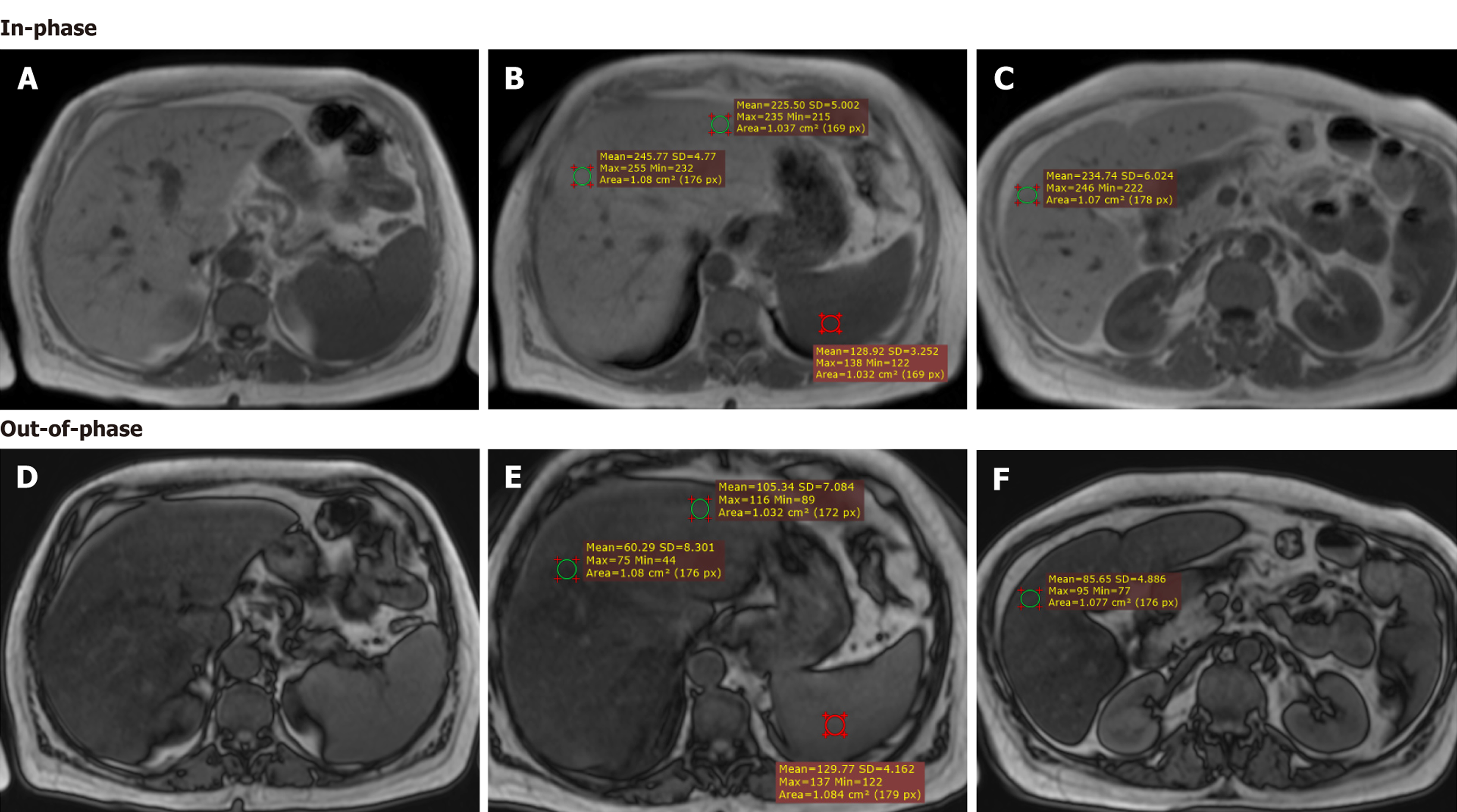
Figure 2 Magnetic resonance imaging of one of our patients exhibiting moderate steatosis.
A-C: Magnetic resonance imaging scan of a female patient, demonstrate axial in-phase; D-F: Out-of-phase sequences showing applied region of interest (ROI’s), calculated hepatic fat percentage (32.2%) and hepatic fat fraction (33%), matching with moderate steatosis.
- Citation: Sholkamy A, El-Meligui A, H Saad E, Amin S, Makram M, Elmansy N, Mousa S. Association between overt, subclinical hypothyroidism and metabolic dysfunction-associated steatotic liver disease using magnetic resonance imaging. World J Hepatol 2026; 18(5): 117441
- URL: https://www.wjgnet.com/1948-5182/full/v18/i5/117441.htm
- DOI: https://dx.doi.org/10.4254/wjh.v18.i5.117441