
















Copyright: ©Author(s) 2026.
World J Hepatol. May 27, 2026; 18(5): 117184
Published online May 27, 2026. doi: 10.4254/wjh.v18.i5.117184
Published online May 27, 2026. doi: 10.4254/wjh.v18.i5.117184
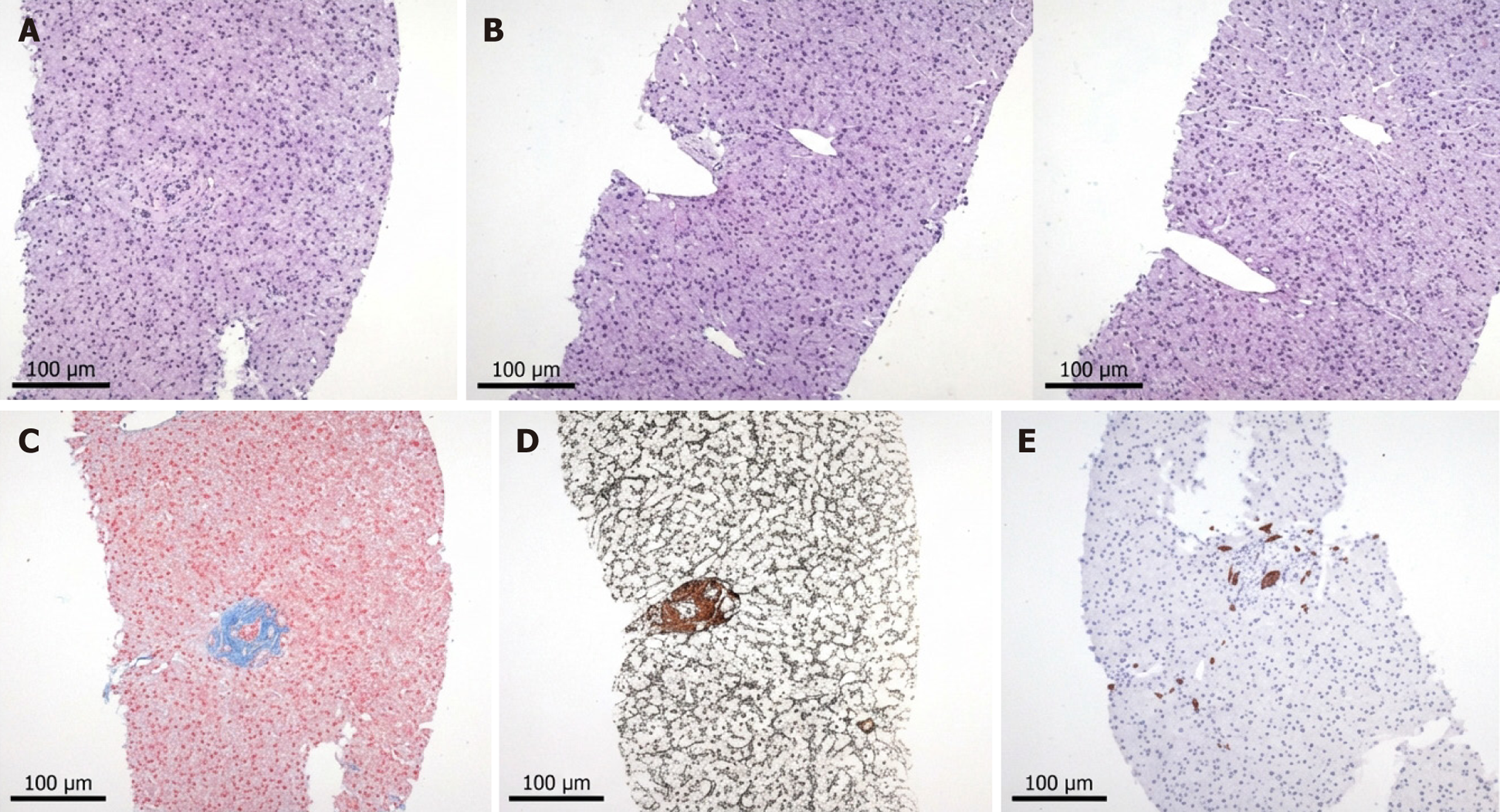
Figure 1 Occlusive portal vein lesions accompanied by nodular regenerative hyperplastic changes.
A: Hepatic sinusoidal lymphocytic infiltration and stromal fibrosis in the confluent zone of the portal vein; B: Portal vein was partially narrowed and occluded, partially tortuous and dilated; C: Peri-sinusoid fibrosis (Masson trichrome); D: Nodular disorganization with hepatocyte plate atrophy (reticulin); E: Immunohistochemical staining showed positivity for CD10, CD38, CK19, and CK7. Scale bars = 100 μm.
- Citation: Xu MJ, Wei X, Huang Y, Lu Y, Sun L, Xie Y, Li MH. Congenital porto-sinusoidal vascular disorder-induced portal hypertension: A comprehensive review based on a classic clinical case. World J Hepatol 2026; 18(5): 117184
- URL: https://www.wjgnet.com/1948-5182/full/v18/i5/117184.htm
- DOI: https://dx.doi.org/10.4254/wjh.v18.i5.117184