
















Copyright: ©Author(s) 2026.
World J Hepatol. May 27, 2026; 18(5): 117157
Published online May 27, 2026. doi: 10.4254/wjh.v18.i5.117157
Published online May 27, 2026. doi: 10.4254/wjh.v18.i5.117157
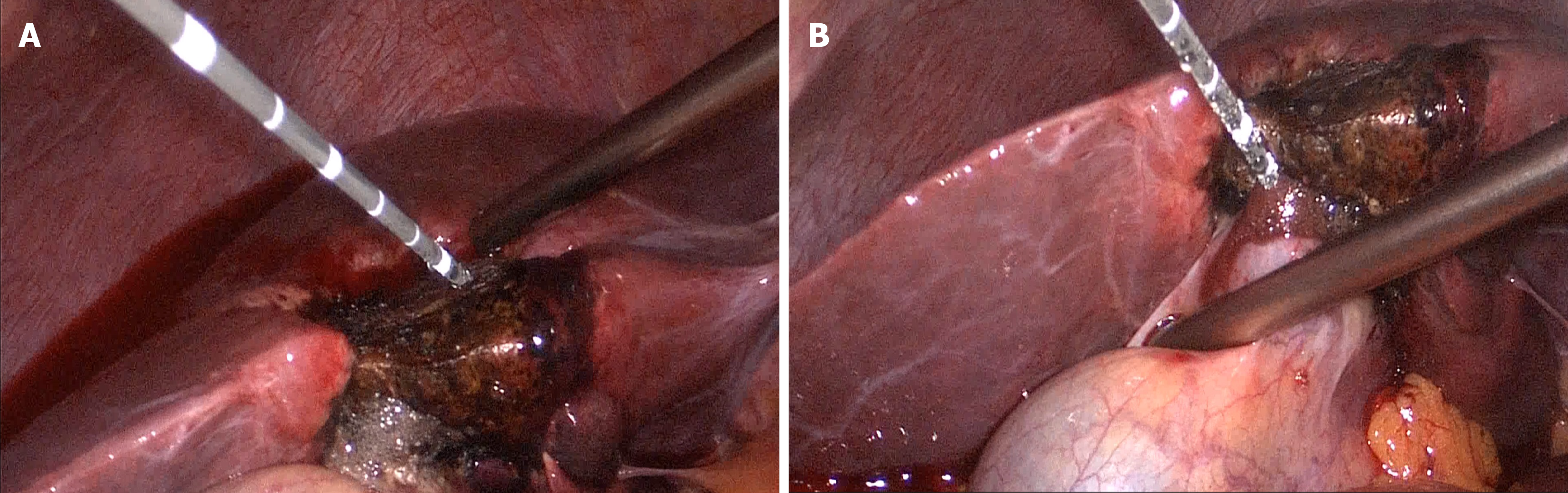
Figure 3 Establishment of the gas-blood outflow channel.
A: Prior to the initiation of each microwave ablation cycle, a gas-blood outflow channel is created using a standard microwave ablation needle. The specific depth of this channel is determined based on the maximum diameter of the hemangioma as measured from preoperative imaging, rather than a fixed length, to ensure tailored channel placement according to individual tumor characteristics (the depth is approximately half of the maximum diameter of the tumor); B: The blood flowing out of the channel, and the thermal oxygen bubbles caused by the heat conduction effect are observed.
- Citation: Wu CL, Chen XC, Zhao W, Hu B, Huan HB, Tu YL, Zhu ZM, Jiang K. Laparoscopic microwave ablation for hepatic hemangiomas with different locations: A comparative study. World J Hepatol 2026; 18(5): 117157
- URL: https://www.wjgnet.com/1948-5182/full/v18/i5/117157.htm
- DOI: https://dx.doi.org/10.4254/wjh.v18.i5.117157