
















Copyright: ©Author(s) 2026.
World J Hepatol. May 27, 2026; 18(5): 117157
Published online May 27, 2026. doi: 10.4254/wjh.v18.i5.117157
Published online May 27, 2026. doi: 10.4254/wjh.v18.i5.117157
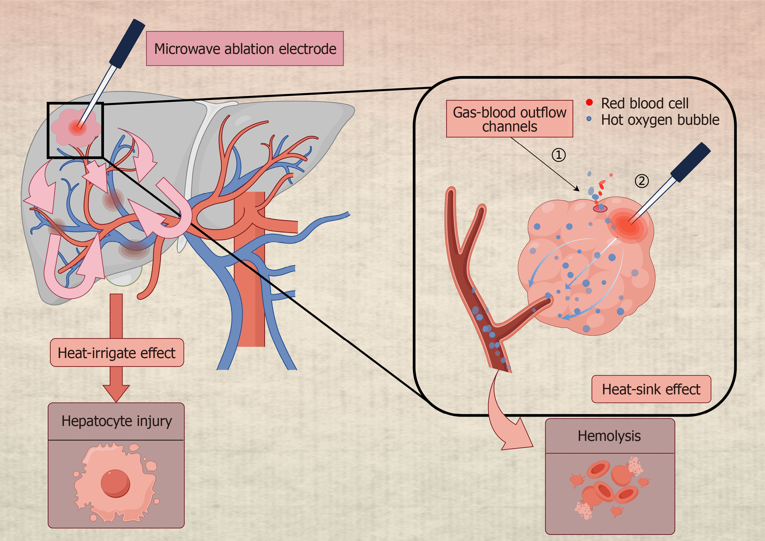
Figure 1 Schematic diagram illustrating microwave ablation of a hepatic hemangioma.
The image shows a microwave electrode inserted into the hemangioma and the generated heat distribution. It depicts heat transfer along the portal vein and heat accumulation within surrounding blood. During the ablation process, the heat from the vessel’s spreads to other liver segments, causing thermal damage to the liver and the accumulation of heat leading to intravascular hemolysis. A channel pre-drilled near the ablation point, known as the gas-blood outflow channel, is conducive to the outflow of hot oxygen bubbles and blood. The image created by FigDraw (Supplementary material).
- Citation: Wu CL, Chen XC, Zhao W, Hu B, Huan HB, Tu YL, Zhu ZM, Jiang K. Laparoscopic microwave ablation for hepatic hemangiomas with different locations: A comparative study. World J Hepatol 2026; 18(5): 117157
- URL: https://www.wjgnet.com/1948-5182/full/v18/i5/117157.htm
- DOI: https://dx.doi.org/10.4254/wjh.v18.i5.117157