
















Copyright: ©Author(s) 2026.
World J Hepatol. Apr 27, 2026; 18(4): 117993
Published online Apr 27, 2026. doi: 10.4254/wjh.v18.i4.117993
Published online Apr 27, 2026. doi: 10.4254/wjh.v18.i4.117993
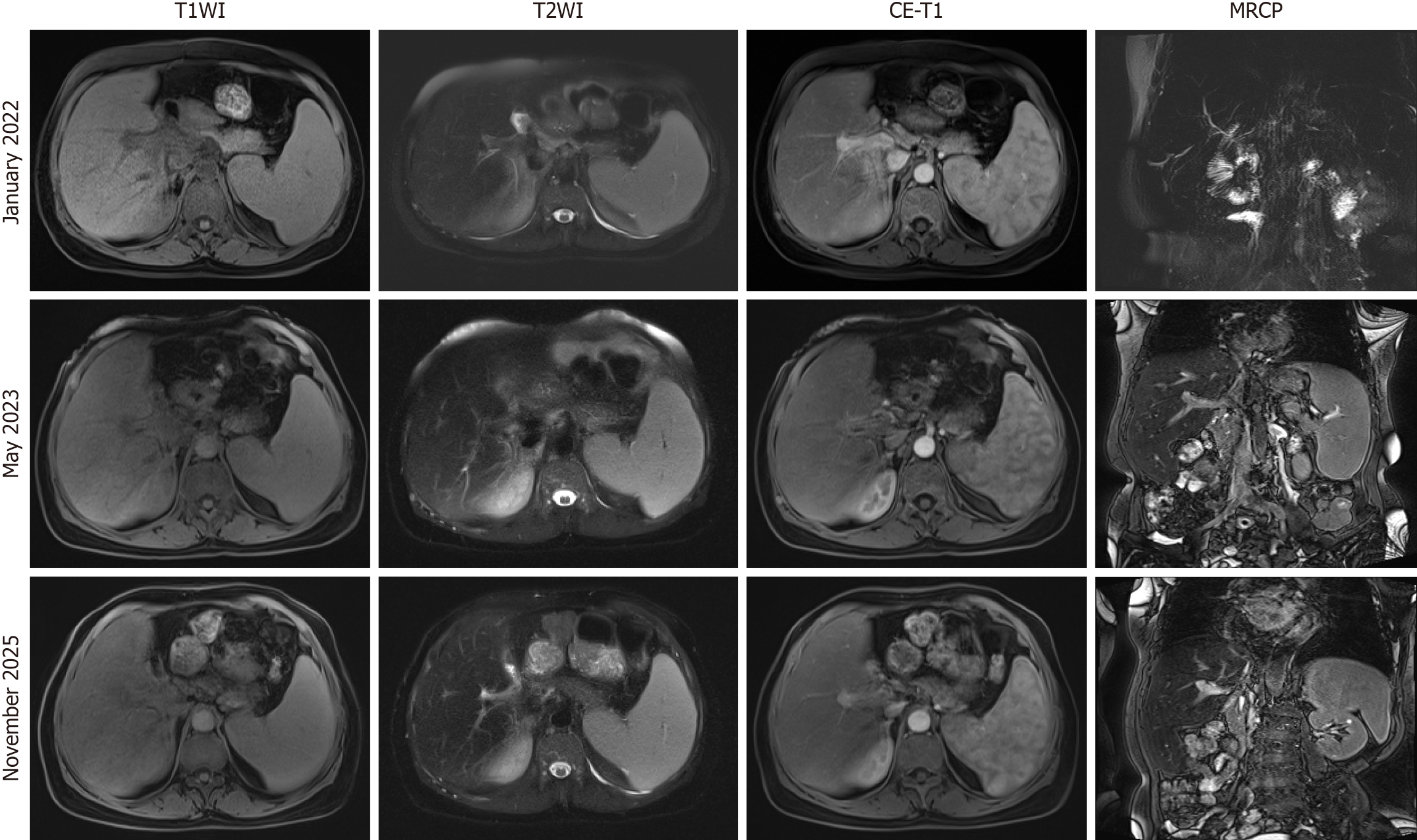
Figure 2 Dynamic evolution of postoperative liver lesions in patients.
Upper abdominal magnetic resonance imaging with magnetic resonance cholangiopancreatography (January 2022, May 2023, November 2025): Imaging revealed post-resection absence of the left hepatic lobe and gallbladder, along with mild intrahepatic biliary dilatation. A rim-enhancing lesion with perilesional hyperperfusion was observed adjacent to the right hepatic hilum, consistent with an inflammatory process. Overall features supported cirrhosis and splenomegaly. Follow-up studies (2023, 2025) showed progressive enlargement of the abnormal perfusion area compared to the initial 2022 study. MRCP: Magnetic resonance cholangiopancreatography.
- Citation: Zhou Y, Cheng QJ, Liang MX, Li SQ, Su YZ, Cai YM, Pan ZQ, Xie YX, Chen YH, He YH. Tumor necrosis factor receptor superfamily member 13B mutation-associated chronic cholangitis with cirrhosis: A case report and review of literature. World J Hepatol 2026; 18(4): 117993
- URL: https://www.wjgnet.com/1948-5182/full/v18/i4/117993.htm
- DOI: https://dx.doi.org/10.4254/wjh.v18.i4.117993