
















Copyright: ©Author(s) 2026.
World J Hepatol. Apr 27, 2026; 18(4): 116710
Published online Apr 27, 2026. doi: 10.4254/wjh.v18.i4.116710
Published online Apr 27, 2026. doi: 10.4254/wjh.v18.i4.116710
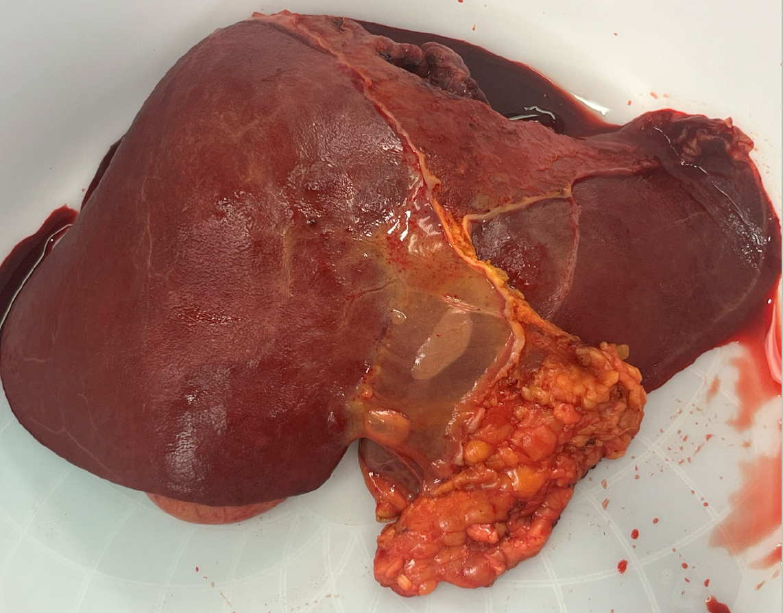
Figure 3 Explanted liver.
Pathology reported extensive hepatocyte necrosis and marked hepatocyte drop-out, involving > 90% of the liver parenchyma (arrows). No viral cytopathic changes. Trichrome stain showed no significant fibrosis. Reticulin stain highlights marked hepatic atrophy. Iron stain was negative for hemosiderosis.
- Citation: Householder S, Brazer N, Ilagan-Ying YC. Acute liver failure linked to OxyShred pre-workout supplement: A case report and review of literature. World J Hepatol 2026; 18(4): 116710
- URL: https://www.wjgnet.com/1948-5182/full/v18/i4/116710.htm
- DOI: https://dx.doi.org/10.4254/wjh.v18.i4.116710