
















Copyright: ©Author(s) 2026.
World J Hepatol. Mar 27, 2026; 18(3): 117040
Published online Mar 27, 2026. doi: 10.4254/wjh.v18.i3.117040
Published online Mar 27, 2026. doi: 10.4254/wjh.v18.i3.117040
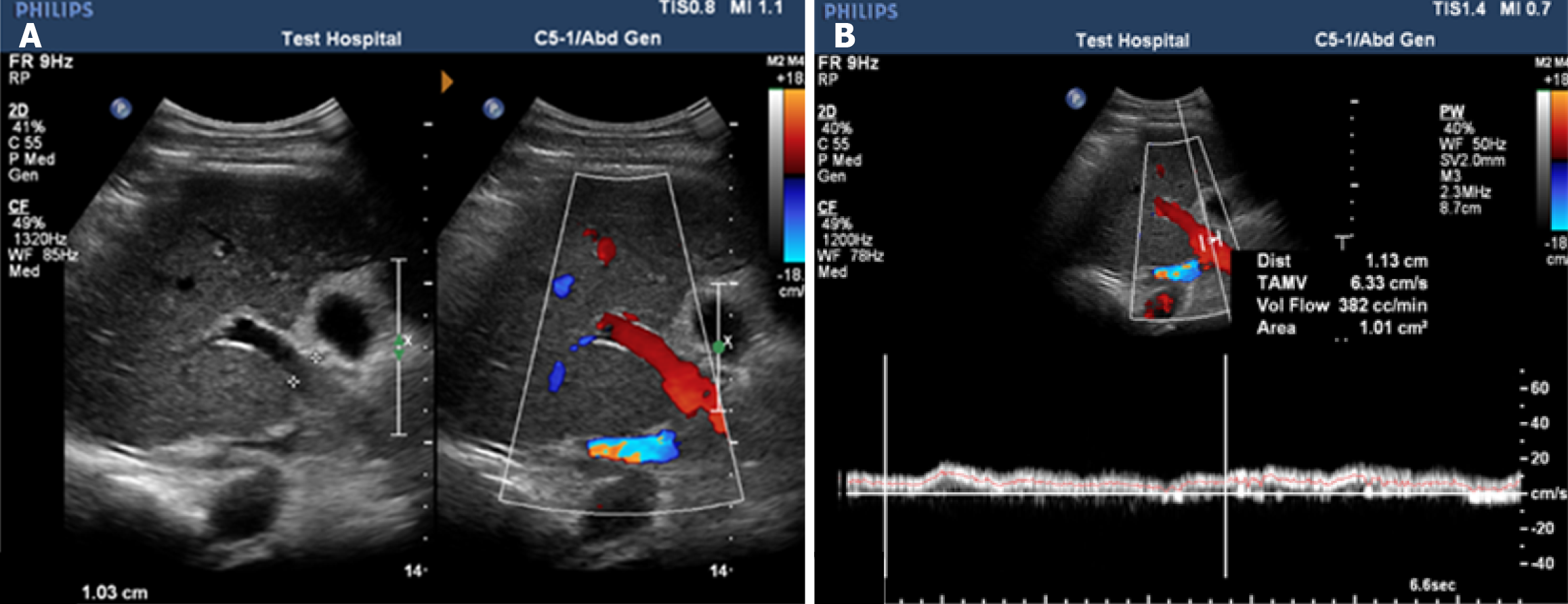
Figure 3 Doppler ultrasonography of the portal vein in a cirrhotic patient with portal hypertension.
A: Dilated portal vein diameter of 11.3 mm; B: Reduced mean flow velocity of 18 cm/second and flow volume of 382 cc/minute.
- Citation: Othman AAA, Elsharkawy L, Saleh MW, Mohamed MM, Elsayed FMA. Multimarker model for non-invasive assessment of variceal bleeding in chronic hepatitis C cirrhotic patients. World J Hepatol 2026; 18(3): 117040
- URL: https://www.wjgnet.com/1948-5182/full/v18/i3/117040.htm
- DOI: https://dx.doi.org/10.4254/wjh.v18.i3.117040