
















©The Author(s) 2026.
World J Hepatol. Feb 27, 2026; 18(2): 113695
Published online Feb 27, 2026. doi: 10.4254/wjh.v18.i2.113695
Published online Feb 27, 2026. doi: 10.4254/wjh.v18.i2.113695
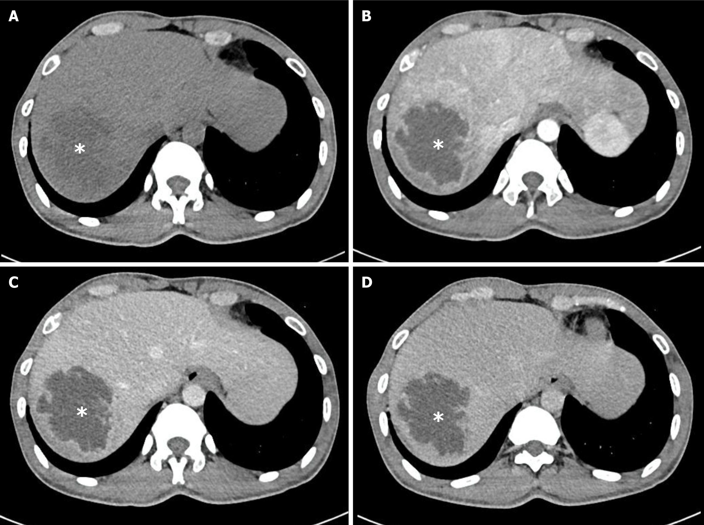
Figure 4 Multiphase computed tomography images of a pyogenic liver abscess caused by Streptococcus species (white asterisks).
The cavity shows slightly irregular margins but homogeneous, non-septated internal architecture with rim enhancement. This contrasts with most Klebsiella pneumoniae liver abscesses, which typically demonstrate heterogeneous internal architecture. A: Non-contrast phase; B: Arterial phase; C: Portal venous phase; D: Delayed phase.
- Citation: Mai-Phan TA, Thai KP, Le KL, Pham TN, Tran MQ, Pham PC, Duong NNQ, Trinh MT, Le NK. Klebsiella pneumoniae as leading cause of pyogenic liver abscess: Three years study in Southern Vietnam. World J Hepatol 2026; 18(2): 113695
- URL: https://www.wjgnet.com/1948-5182/full/v18/i2/113695.htm
- DOI: https://dx.doi.org/10.4254/wjh.v18.i2.113695