
















©The Author(s) 2026.
World J Hepatol. Feb 27, 2026; 18(2): 113686
Published online Feb 27, 2026. doi: 10.4254/wjh.v18.i2.113686
Published online Feb 27, 2026. doi: 10.4254/wjh.v18.i2.113686
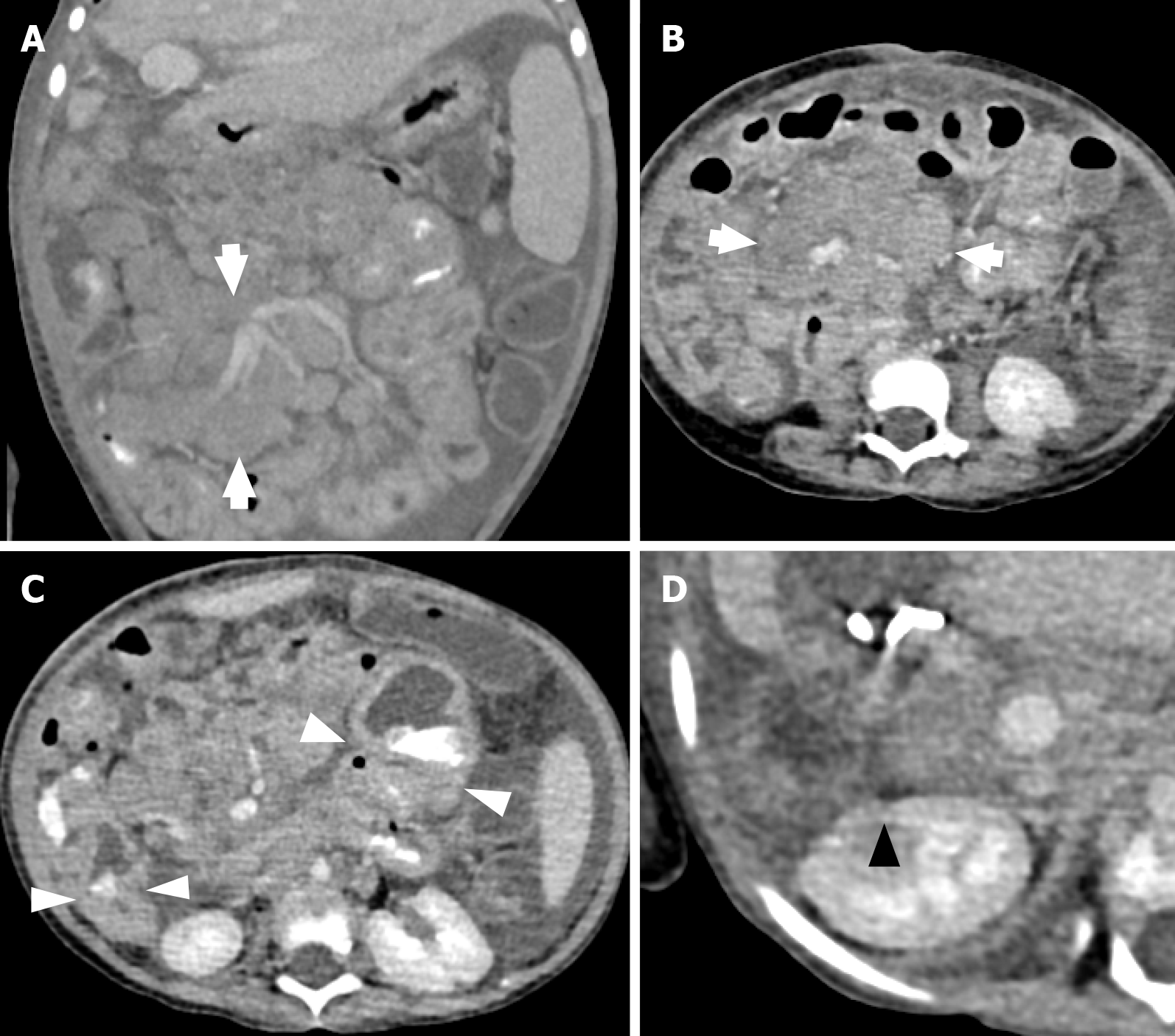
Figure 5 Computed tomography images.
A and B: Computed tomography images of a 34-month-old girl who presented with pallor and mucous diarrhea after liver transplantation showed large, matted mesenteric lymph nodes (white arrows); C: A few small bowel loops with focal wall thickening (white arrowheads); D: A hypoenhancing lesion in the right kidney (black triangle).
- Citation: Onpoaree N, Leelakanok N, Sanpavat A, Sintusek P. Epstein-Barr virus infection in children with liver transplantation. World J Hepatol 2026; 18(2): 113686
- URL: https://www.wjgnet.com/1948-5182/full/v18/i2/113686.htm
- DOI: https://dx.doi.org/10.4254/wjh.v18.i2.113686