
















©The Author(s) 2026.
World J Hepatol. Feb 27, 2026; 18(2): 113004
Published online Feb 27, 2026. doi: 10.4254/wjh.v18.i2.113004
Published online Feb 27, 2026. doi: 10.4254/wjh.v18.i2.113004
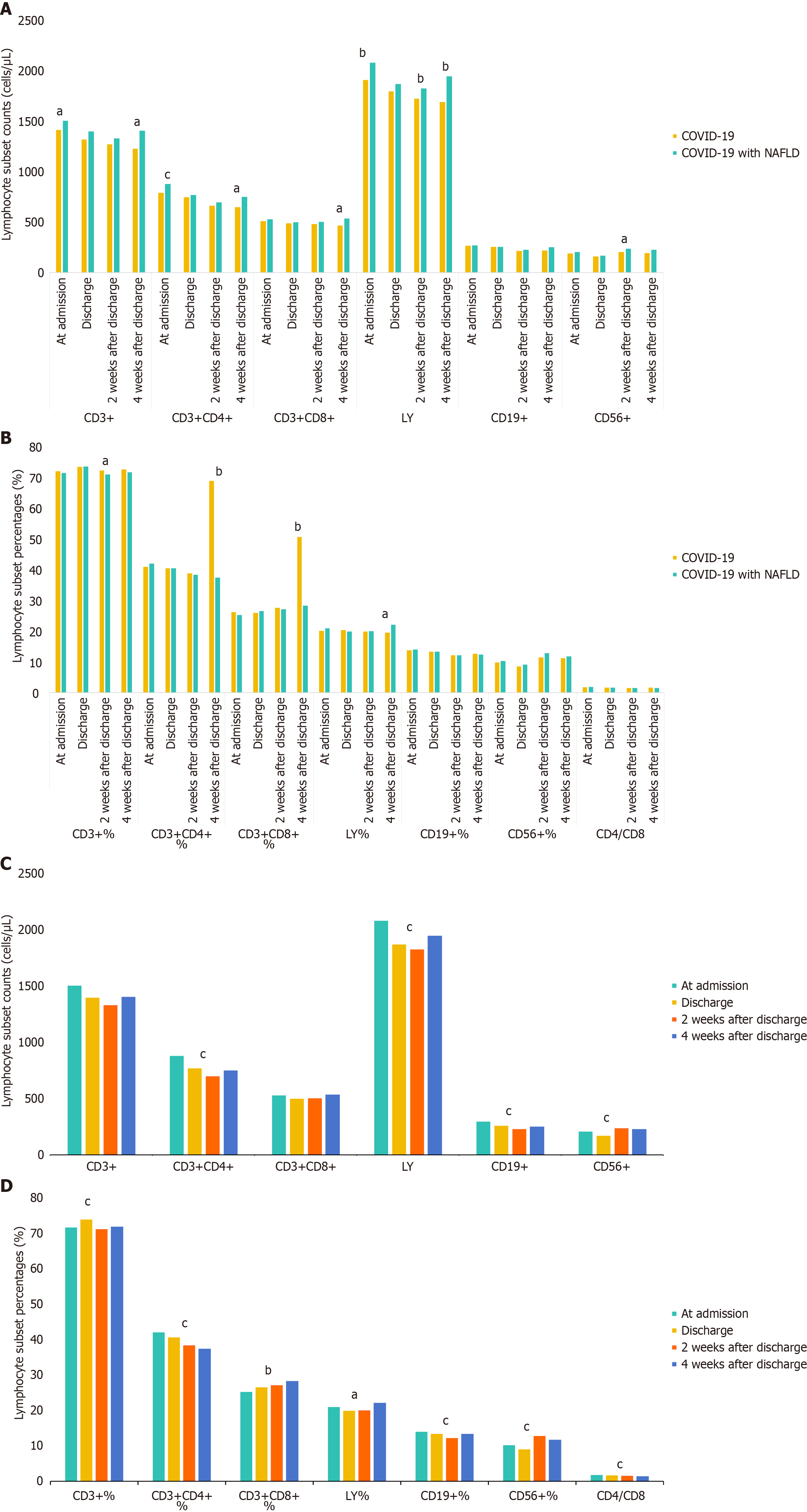
Figure 2 Analysis of lymphocyte subsets and their percentages in coronavirus disease 2019 patients with non-alcoholic fatty liver disease at admission, discharge, and 2 weeks and 4 weeks post-discharge after adjustment for comorbidities.
A and B: Comparison between coronavirus disease 2019 (COVID-19) patients with non-alcoholic fatty liver disease and those without; C and D: Comparison across different time points in COVID-19 patients with non-alcoholic fatty liver disease. aP < 0.05; bP < 0.01; cP < 0.001. COVID-19: Coronavirus disease 2019; LY: Lymphocyte counts.
- Citation: Yan P, Li R, Yuan XY, Wang Y, Lan LJ, Yu XP, Liu DF. Dynamic immune response and its influencing factors in COVID-19 patients with non-alcoholic fatty liver disease: A cohort study. World J Hepatol 2026; 18(2): 113004
- URL: https://www.wjgnet.com/1948-5182/full/v18/i2/113004.htm
- DOI: https://dx.doi.org/10.4254/wjh.v18.i2.113004