
















Copyright: ©Author(s) 2026.
World J Hepatol. Oct 27, 2026; 18(10): 121142
Published online Oct 27, 2026. doi: 10.4254/wjh.121142
Published online Oct 27, 2026. doi: 10.4254/wjh.121142
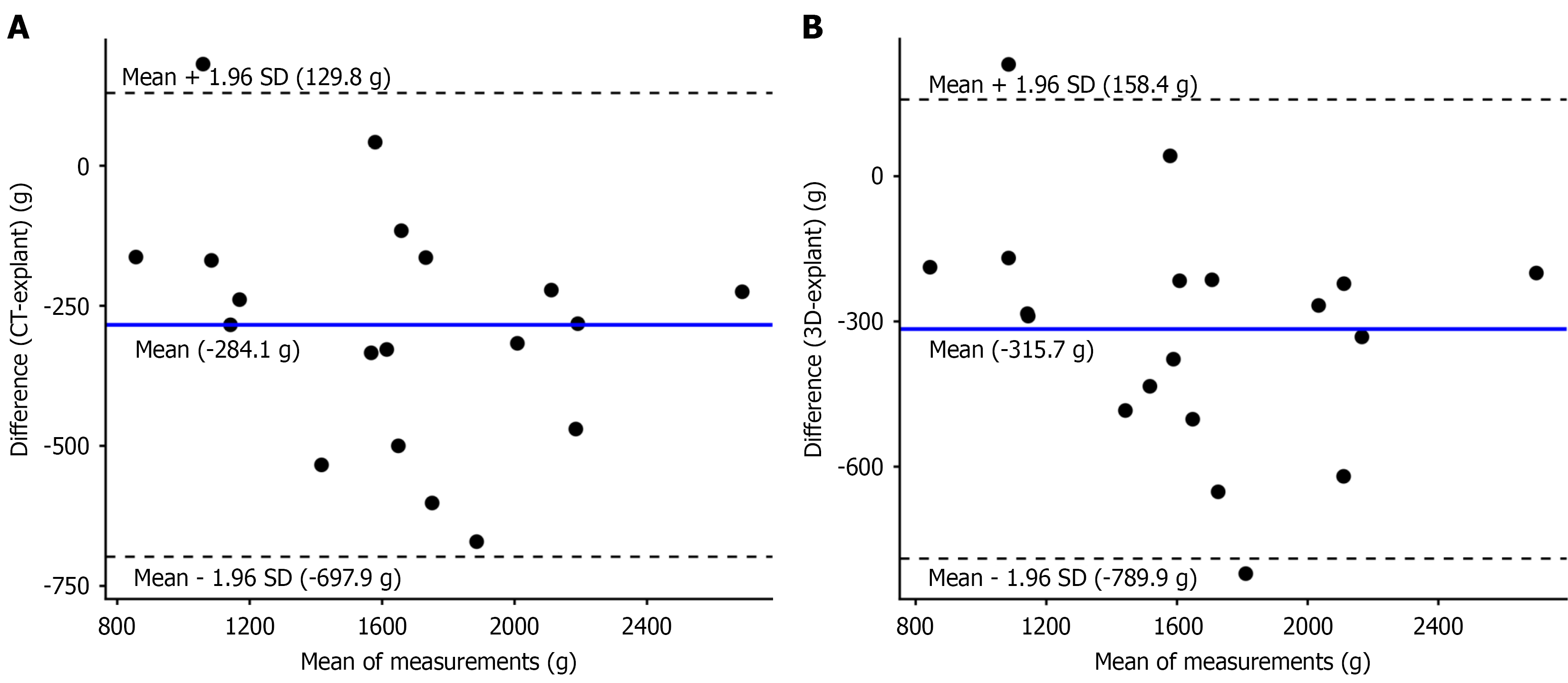
Figure 2 Agreement between imaging-based graft size estimates and explant liver mass.
Bland-Altman plots showing the differences between imaging-based graft size estimates and explant liver mass plotted against the mean of the measurements. A: Computed tomography volumetry vs explant liver mass; B: Three-dimensional surface-derived volumetry vs explant liver mass. Dashed lines indicate the mean bias and 95% limits of agreement. Values are expressed in grams (g). CT: Computed tomography; 3D: Three-dimensional.
- Citation: Jaya LOI, Rykowski P, Bartkowiak M, Kruk E, Chodurska M, Lewandowski Z, Krasnodębski M, Grąt M. Computed tomography volumetry vs three-dimensional surface scanning for liver graft size estimation using explant liver mass. World J Hepatol 2026; 18(10): 121142
- URL: https://www.wjgnet.com/1948-5182/full/v18/i10/121142.htm
- DOI: https://dx.doi.org/10.4254/wjh.121142