
















©The Author(s) 2026.
World J Hepatol. Jan 27, 2026; 18(1): 114542
Published online Jan 27, 2026. doi: 10.4254/wjh.v18.i1.114542
Published online Jan 27, 2026. doi: 10.4254/wjh.v18.i1.114542
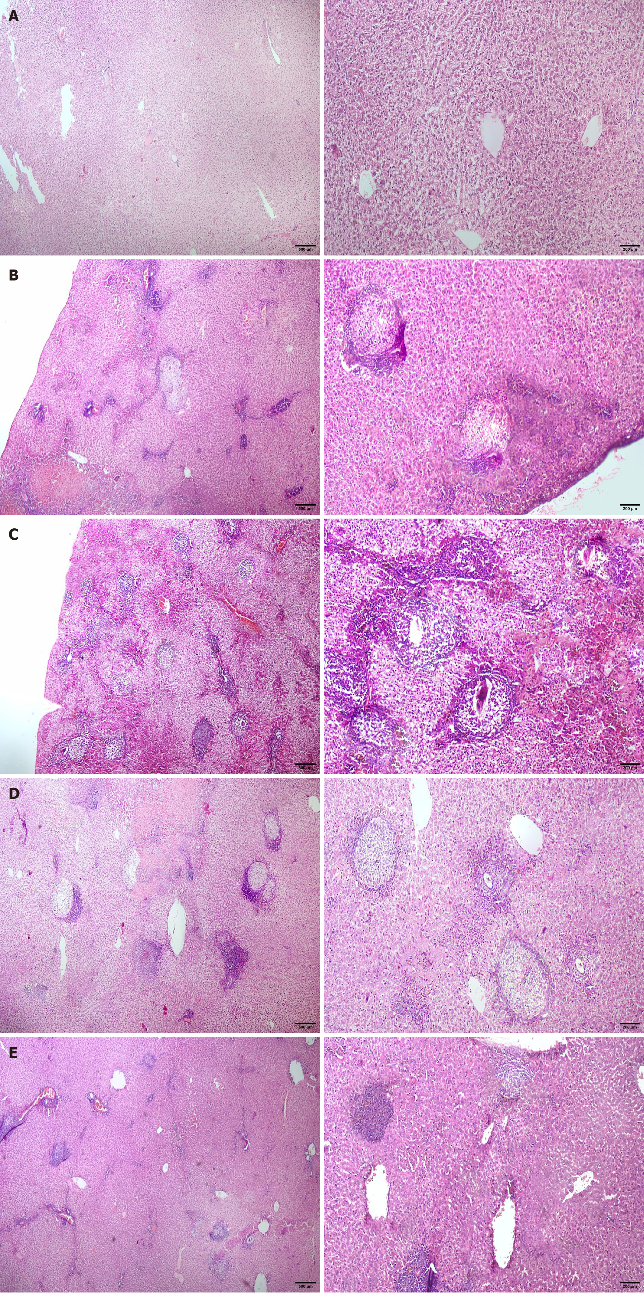
Figure 2 Hematoxylin and eosin micrograph of liver sections.
A and B: Normal hepatic structure with hepatocytes arranged in strands alternating with blood sinusoids forming a network around central veins. No neuroinflammatory injury or granulomas were detected. Control negative group I (A); control negative Ziziphus spina-christi leaf extract (ZSCLE)-treated group II (B); C: Group III with infected Bovine leukemia virus (BLV), showing multiple bilharzial granulomas formed of bilharzial ova, eosinophils, epithelioid cells, and fibrous capsule; D: Infected BLV and ZSCLE-treated group IV, showing moderate granuloma formation accompanied by inflammatory reaction; E: Infected BLV and ZSCLE-treated + 50% resected group V, showing few granulomas with remaining focal inflammatory cell infiltration and congestion.
- Citation: Alghamdi TH. Enhanced surgical tolerance of schistosomiasis-induced fibrotic liver following treatment with Ziziphus plant extract. World J Hepatol 2026; 18(1): 114542
- URL: https://www.wjgnet.com/1948-5182/full/v18/i1/114542.htm
- DOI: https://dx.doi.org/10.4254/wjh.v18.i1.114542