
















©The Author(s) 2026.
World J Hepatol. Jan 27, 2026; 18(1): 114542
Published online Jan 27, 2026. doi: 10.4254/wjh.v18.i1.114542
Published online Jan 27, 2026. doi: 10.4254/wjh.v18.i1.114542
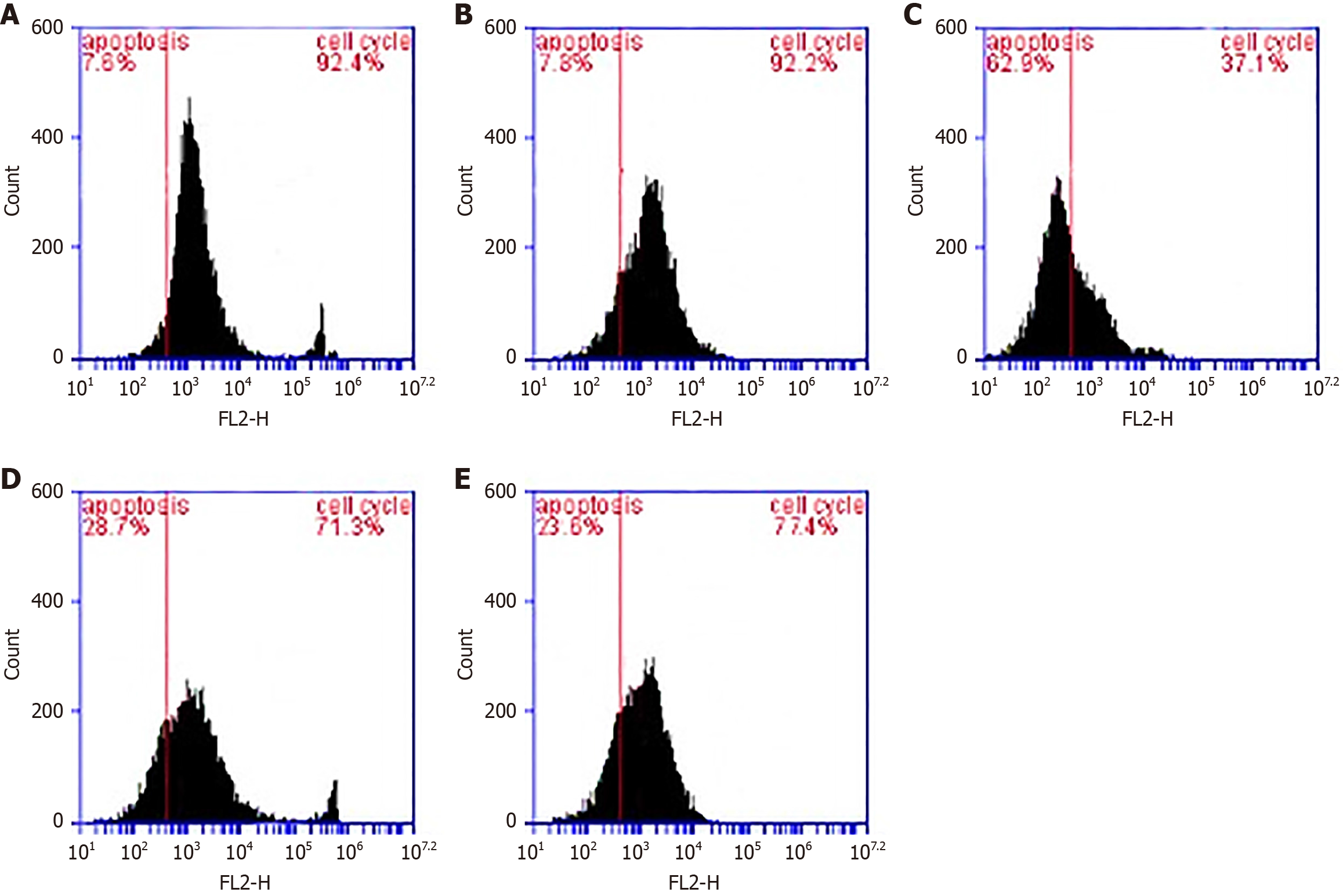
Figure 1 Apoptosis analysis.
A and B: Liver cell damage, represented by apoptosis, was normal in group I and I group II, respectively; C: A higher percentage of liver damage (62.9%) was observed in group III hamsters with infected Bovine leukemia virus; D and E: A marked decline in apoptosis (28.7% and 23.6%, respectively) in the groups that received Ziziphus spina-christi leaf extract (group IV) and Ziziphus spina-christi leaf extract + 50% liver resection (group V). Apoptosis is identified by the dark, black-stained area upon propidium iodide staining, which labels DNA; the stained area denotes marked DNA fragmentation. FL2-H: Fluorescence channel 2 height.
- Citation: Alghamdi TH. Enhanced surgical tolerance of schistosomiasis-induced fibrotic liver following treatment with Ziziphus plant extract. World J Hepatol 2026; 18(1): 114542
- URL: https://www.wjgnet.com/1948-5182/full/v18/i1/114542.htm
- DOI: https://dx.doi.org/10.4254/wjh.v18.i1.114542