
















©The Author(s) 2026.
World J Hepatol. Jan 27, 2026; 18(1): 112821
Published online Jan 27, 2026. doi: 10.4254/wjh.v18.i1.112821
Published online Jan 27, 2026. doi: 10.4254/wjh.v18.i1.112821
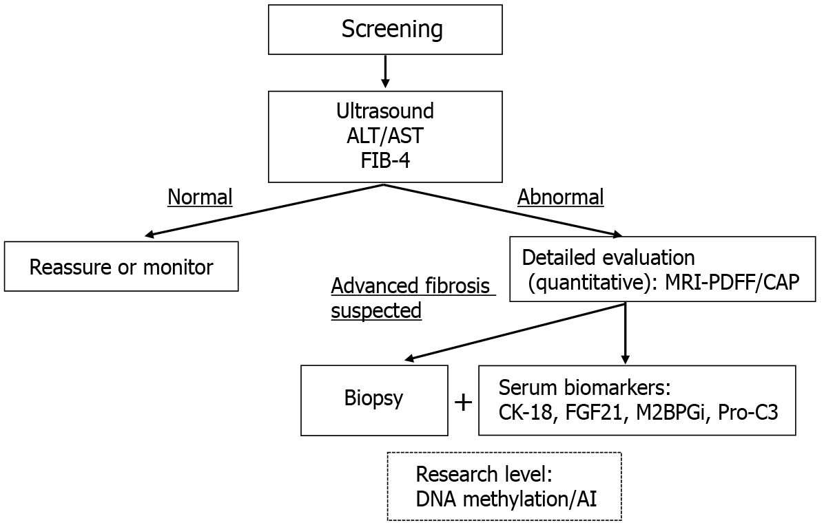
Figure 2 Diagnostic algorithm for hepatic steatosis evaluation.
This flowchart illustrates a stepwise approach to diagnosing hepatic steatosis based on clinical context. Screening typically begins with noninvasive methods such as ultrasound and liver enzyme assessment (alanine aminotransferase/aspartate aminotransferase) along with simple fibrosis scores (e.g., fibrosis-4). Patients with abnormal results proceed to more quantitative assessments using magnetic resonance imaging-proton density fat fraction or controlled attenuation parameter. In cases where advanced fibrosis is suspected, both serum biomarkers (e.g., cytokeratin-18, fibroblast growth factor 21, Mac-2 binding protein glycosylation isomer, type III procollagen peptide) and liver biopsy may be considered. Biopsy remains the gold standard for histological confirmation. Emerging research-level diagnostic methods include DNA methylation profiling and artificial intelligence-based tools. ALT: Alanine aminotransferase; AST: Aspartate aminotransferase; FIB-4: Fibrosis-4; MRI-PDFF: Magnetic resonance imaging-proton density fat fraction; CAP: Controlled attenuation parameter; CK-18: Cytokeratin-18; FGF21: Fibroblast growth factor 21; M2BPGi: Mac-2 binding protein glycosylation isomer; Pro-C3: Type III procollagen peptide; AI: Artificial intelligence.
- Citation: Moriyama K. Evaluation methods of hepatic steatosis: From conventional techniques to emerging biomarkers. World J Hepatol 2026; 18(1): 112821
- URL: https://www.wjgnet.com/1948-5182/full/v18/i1/112821.htm
- DOI: https://dx.doi.org/10.4254/wjh.v18.i1.112821