
















©The Author(s) 2025.
World J Hepatol. Sep 27, 2025; 17(9): 107705
Published online Sep 27, 2025. doi: 10.4254/wjh.v17.i9.107705
Published online Sep 27, 2025. doi: 10.4254/wjh.v17.i9.107705
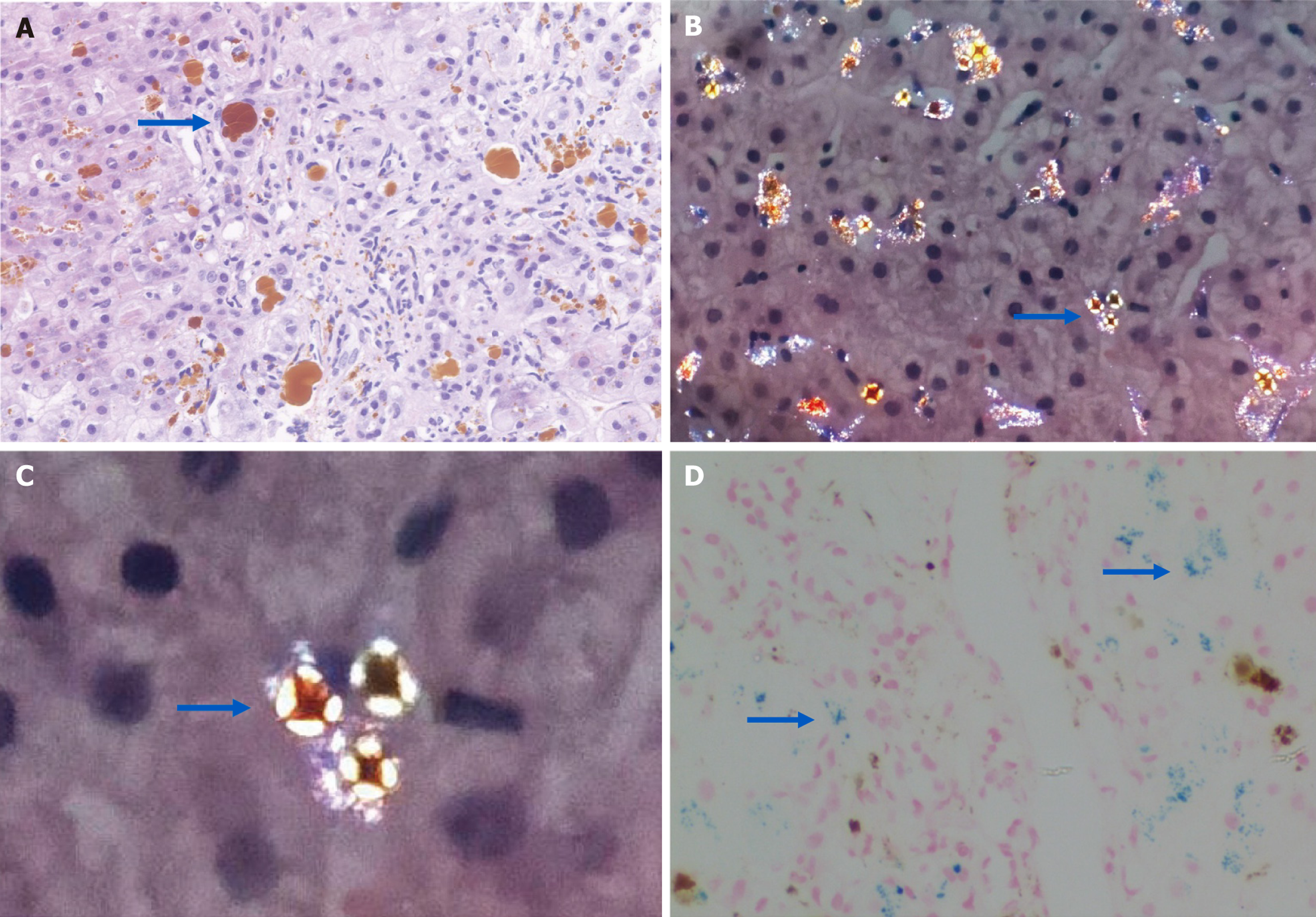
Figure 3 Histopathological features of protoporphyria-associated liver injury.
A: Hematoxylin and eosin staining shows brown-yellow protoporphyrin deposits within canaliculi and hepatocytes; B and C: Polarized light microscopy reveals Maltese cross-shaped birefringent crystals (blue arrows), indicative of protoporphyrin aggregation; D: Prussian blue staining highlights granular iron deposits (blue arrows).
- Citation: Zeng T, Huang SY, Chen JN, Pang JH, Chong YT, Li XH. Pathogenesis and clinical management of liver damage in porphyrias: Mechanisms and therapeutic approaches. World J Hepatol 2025; 17(9): 107705
- URL: https://www.wjgnet.com/1948-5182/full/v17/i9/107705.htm
- DOI: https://dx.doi.org/10.4254/wjh.v17.i9.107705