Copyright
©The Author(s) 2025.
World J Hepatol. Aug 27, 2025; 17(8): 107873
Published online Aug 27, 2025. doi: 10.4254/wjh.v17.i8.107873
Published online Aug 27, 2025. doi: 10.4254/wjh.v17.i8.107873
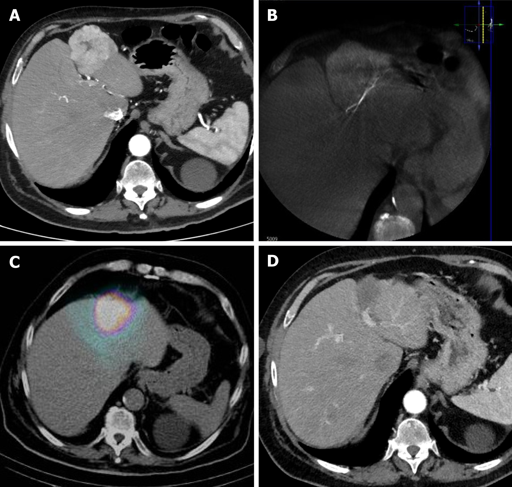
Figure 3 69-year-old man with hepatocellular carcinoma unfit for surgery due to comorbidities.
A: Computed tomography (CT) showing hepatocellular carcinoma nodule (5-6 cm) located in S2; B: Cone beam CT demonstrating superselective catheterization of the segmental S2 artery with perfused nodule; C: Single-photon emission tomography combined with CT showing 99mTc-MAA uptake into S2; D: 12-months CT follow-up scan showing complete nodule necrosis with no contrast enhancement and atrophy of the treated liver lobe (radiation segmentectomy).
- Citation: Cortese F, Anagnostopoulos F, Bazzocchi MV, Caringi S, Pisani AR, Renzulli M, Paraskevopoulos I, Laera L, Surgo A, Spiliopoulos S, Memeo R, Inchingolo R. Modern approach to hepatocellular carcinoma treatment. World J Hepatol 2025; 17(8): 107873
- URL: https://www.wjgnet.com/1948-5182/full/v17/i8/107873.htm
- DOI: https://dx.doi.org/10.4254/wjh.v17.i8.107873


