
















©The Author(s) 2025.
World J Hepatol. Aug 27, 2025; 17(8): 107679
Published online Aug 27, 2025. doi: 10.4254/wjh.v17.i8.107679
Published online Aug 27, 2025. doi: 10.4254/wjh.v17.i8.107679
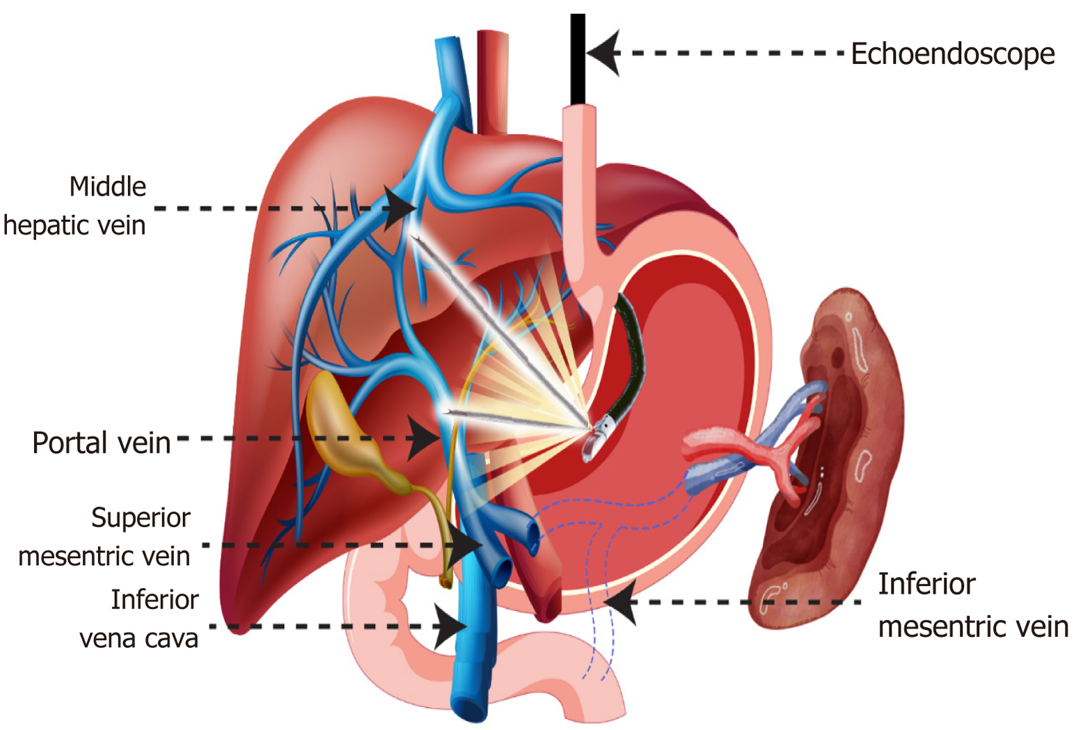
Figure 2 Technique of doing endoscopic ultrasound guided portal pressure gradient measurement.
The hepatic vein (HV) is usually punctured first followed by the portal vein (PV). Middle HV and umbilicated portion of the left PV are the preferred site of puncture respectively due to larger calibre and better alignment using a 22G or 25G fine needle aspiration (FNA) needle. The FNA needle is attached to the non-compressible tubing which is then attached to either compact manometer or the conventional electro-cardiogram monitor depending upon availability. With patient in supine position and pressure transducer kept at the level of mid-axillary line, three sequential readings are taken for each vessel for 60 seconds and mean calculated.
- Citation: Singla N, Shantan V, Saraswat A, Singh AP. Advances in portal pressure measurement: Endoscopic techniques, challenges, and implications for liver transplantation. World J Hepatol 2025; 17(8): 107679
- URL: https://www.wjgnet.com/1948-5182/full/v17/i8/107679.htm
- DOI: https://dx.doi.org/10.4254/wjh.v17.i8.107679