
















©The Author(s) 2025.
World J Hepatol. Aug 27, 2025; 17(8): 107041
Published online Aug 27, 2025. doi: 10.4254/wjh.v17.i8.107041
Published online Aug 27, 2025. doi: 10.4254/wjh.v17.i8.107041
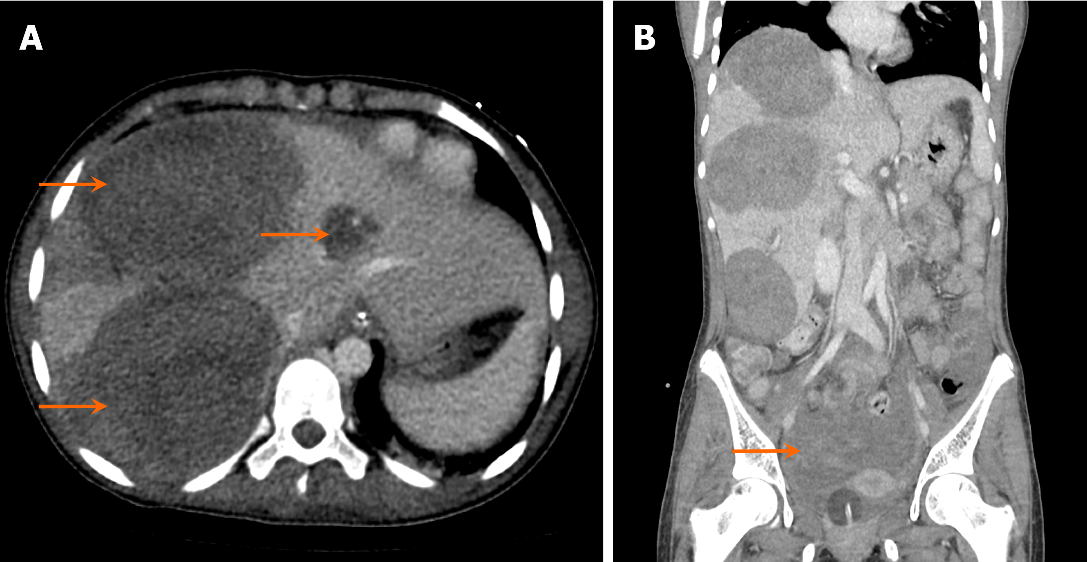
Figure 24 A 14-year-old girl, who was a known case of left ovarian mixed germ cell tumor (95% dysgerminoma), presented with widespread liver metastasis and hemoperitoneum.
A: Axial computed tomography (CT) image; B: Coronal CT image. Images show numerous large liver lesions (arrows in A) with high-density ascites suggesting tumor rupture (arrow in B).
- Citation: Shahid M, Hilal K, Khan M, Ejaz ZH, Altaf S, Islam S, Khandwala K. Imaging insights into pediatric liver masses: A comprehensive minireview for hepatology practice. World J Hepatol 2025; 17(8): 107041
- URL: https://www.wjgnet.com/1948-5182/full/v17/i8/107041.htm
- DOI: https://dx.doi.org/10.4254/wjh.v17.i8.107041