Copyright
©The Author(s) 2025.
World J Hepatol. Aug 27, 2025; 17(8): 107041
Published online Aug 27, 2025. doi: 10.4254/wjh.v17.i8.107041
Published online Aug 27, 2025. doi: 10.4254/wjh.v17.i8.107041
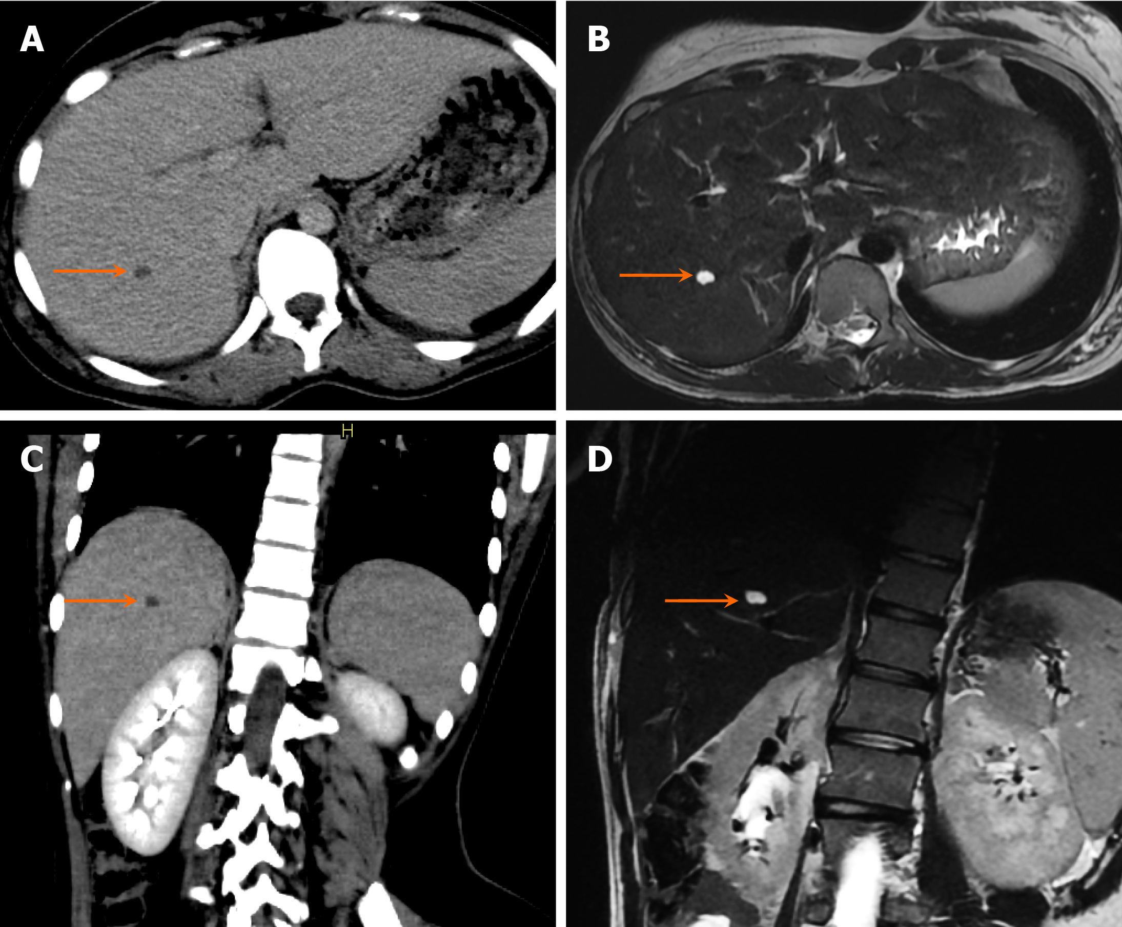
Figure 14 Simple hepatic cyst in a 16-year-old girl who presented with complaints of abdominal pain.
A and B: Axial computed tomography (CT) and T2-weighted magnetic resonance (MR) images; C and D: Coronal CT and T2-weighted MR images. CT images with corresponding T2-weighted images from magnetic resonance imaging show small hypodense and T2 hyperintense lesions in the right lobe of the liver, respectively, involving segment VII, which has thin walls without internal debris or septations, and no apparent biliary communication, indicating simple hepatic cysts (arrows).
- Citation: Shahid M, Hilal K, Khan M, Ejaz ZH, Altaf S, Islam S, Khandwala K. Imaging insights into pediatric liver masses: A comprehensive minireview for hepatology practice. World J Hepatol 2025; 17(8): 107041
- URL: https://www.wjgnet.com/1948-5182/full/v17/i8/107041.htm
- DOI: https://dx.doi.org/10.4254/wjh.v17.i8.107041


