Copyright
©The Author(s) 2025.
World J Hepatol. Aug 27, 2025; 17(8): 107041
Published online Aug 27, 2025. doi: 10.4254/wjh.v17.i8.107041
Published online Aug 27, 2025. doi: 10.4254/wjh.v17.i8.107041
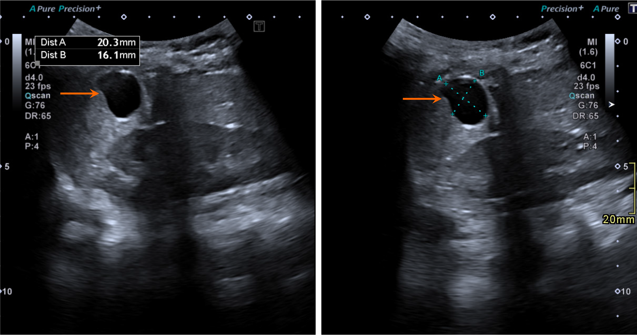
Figure 13 Simple hepatic cyst in a 7-year-old girl who underwent grayscale ultrasound for nonspecific abdominal pain.
An anechoic structure (arrow) is seen in the right lobe of the liver involving segment VI, without any internal debris or septae, which is consistent with a simple hepatic cyst.
- Citation: Shahid M, Hilal K, Khan M, Ejaz ZH, Altaf S, Islam S, Khandwala K. Imaging insights into pediatric liver masses: A comprehensive minireview for hepatology practice. World J Hepatol 2025; 17(8): 107041
- URL: https://www.wjgnet.com/1948-5182/full/v17/i8/107041.htm
- DOI: https://dx.doi.org/10.4254/wjh.v17.i8.107041


