Copyright
©The Author(s) 2025.
World J Hepatol. Aug 27, 2025; 17(8): 107041
Published online Aug 27, 2025. doi: 10.4254/wjh.v17.i8.107041
Published online Aug 27, 2025. doi: 10.4254/wjh.v17.i8.107041
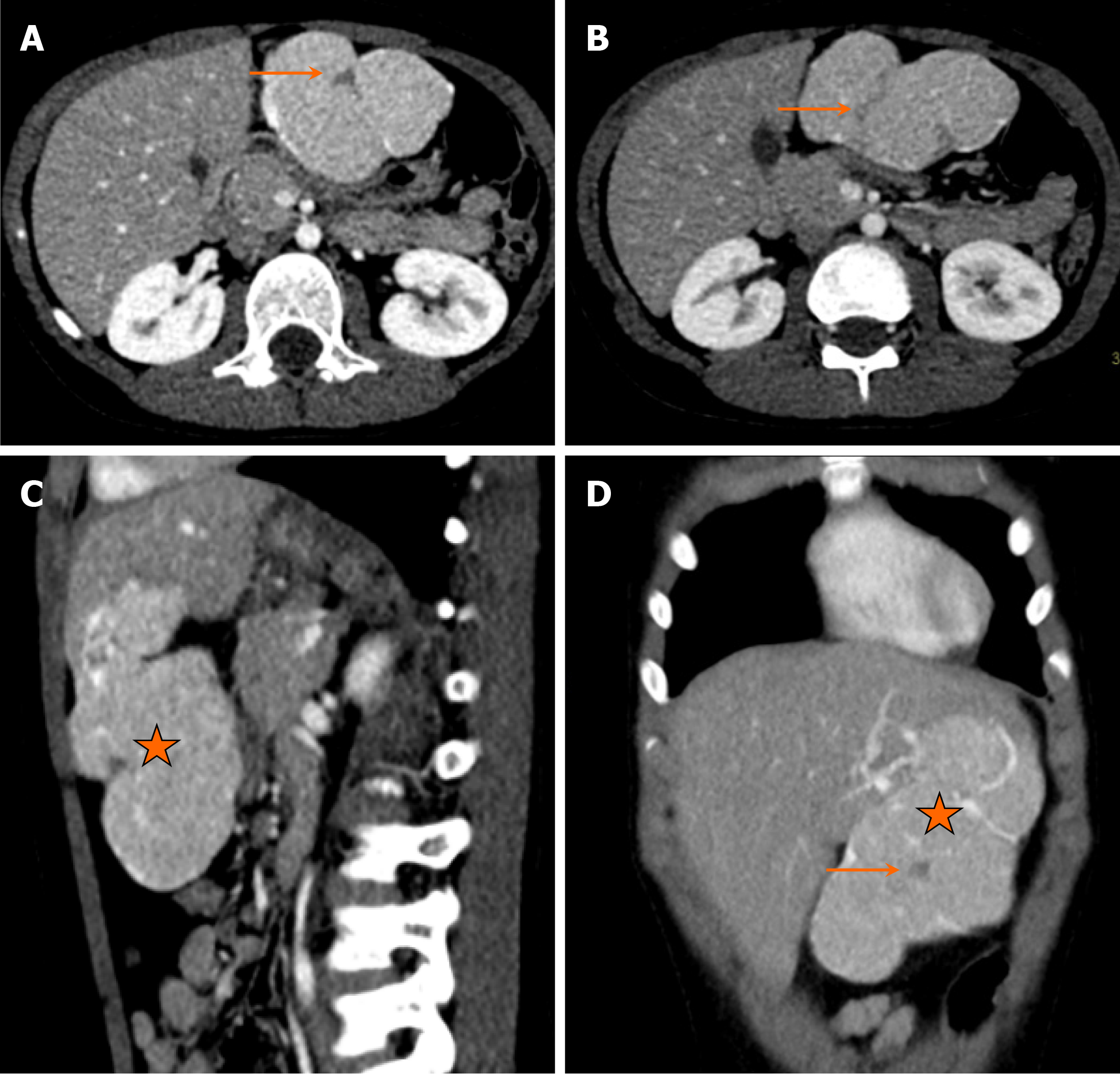
Figure 11 Focal nodular hyperplasia in a 9-year-old boy with complaints of progressively worsening abdominal pain for one year and loose stools for five months.
A and B: Axial images; C: Sagittal image; D: Coronal image. Images from computed tomography scan show a large, well-defined, exophytic, intensely enhancing hepatic lesion (asterisk in C and D) arising from the left hepatic lobe, involving segments II and III, with a central hypodense scar (arrows in A, B and D).
- Citation: Shahid M, Hilal K, Khan M, Ejaz ZH, Altaf S, Islam S, Khandwala K. Imaging insights into pediatric liver masses: A comprehensive minireview for hepatology practice. World J Hepatol 2025; 17(8): 107041
- URL: https://www.wjgnet.com/1948-5182/full/v17/i8/107041.htm
- DOI: https://dx.doi.org/10.4254/wjh.v17.i8.107041


