
















©The Author(s) 2025.
World J Hepatol. Aug 27, 2025; 17(8): 107041
Published online Aug 27, 2025. doi: 10.4254/wjh.v17.i8.107041
Published online Aug 27, 2025. doi: 10.4254/wjh.v17.i8.107041
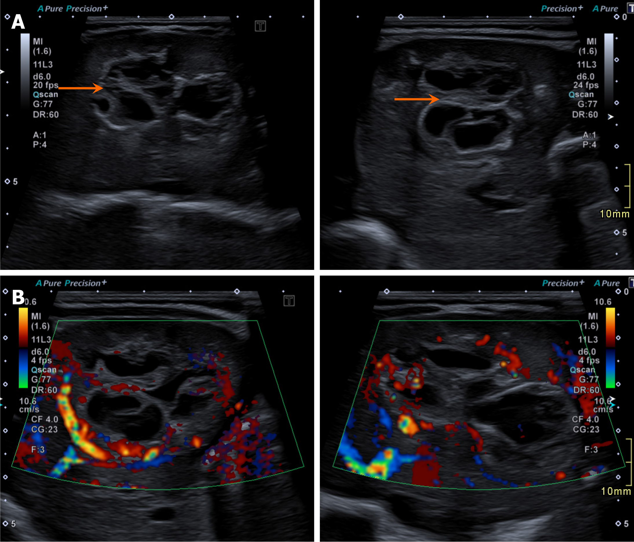
Figure 8 Mesenchymal hamartoma of the liver in a 1-day-old baby boy with a history of a multicystic echogenic structure in the right side of the abdomen on antenatal scan.
A: Grayscale ultrasound (US) image; B: Color Doppler US image. Images show a large lobulated heterogeneous multiseptated cystic lesion in the liver interspersed with solid component (arrows in A) with some internal vascularity in B.
- Citation: Shahid M, Hilal K, Khan M, Ejaz ZH, Altaf S, Islam S, Khandwala K. Imaging insights into pediatric liver masses: A comprehensive minireview for hepatology practice. World J Hepatol 2025; 17(8): 107041
- URL: https://www.wjgnet.com/1948-5182/full/v17/i8/107041.htm
- DOI: https://dx.doi.org/10.4254/wjh.v17.i8.107041