Copyright
©The Author(s) 2025.
World J Hepatol. Aug 27, 2025; 17(8): 107041
Published online Aug 27, 2025. doi: 10.4254/wjh.v17.i8.107041
Published online Aug 27, 2025. doi: 10.4254/wjh.v17.i8.107041
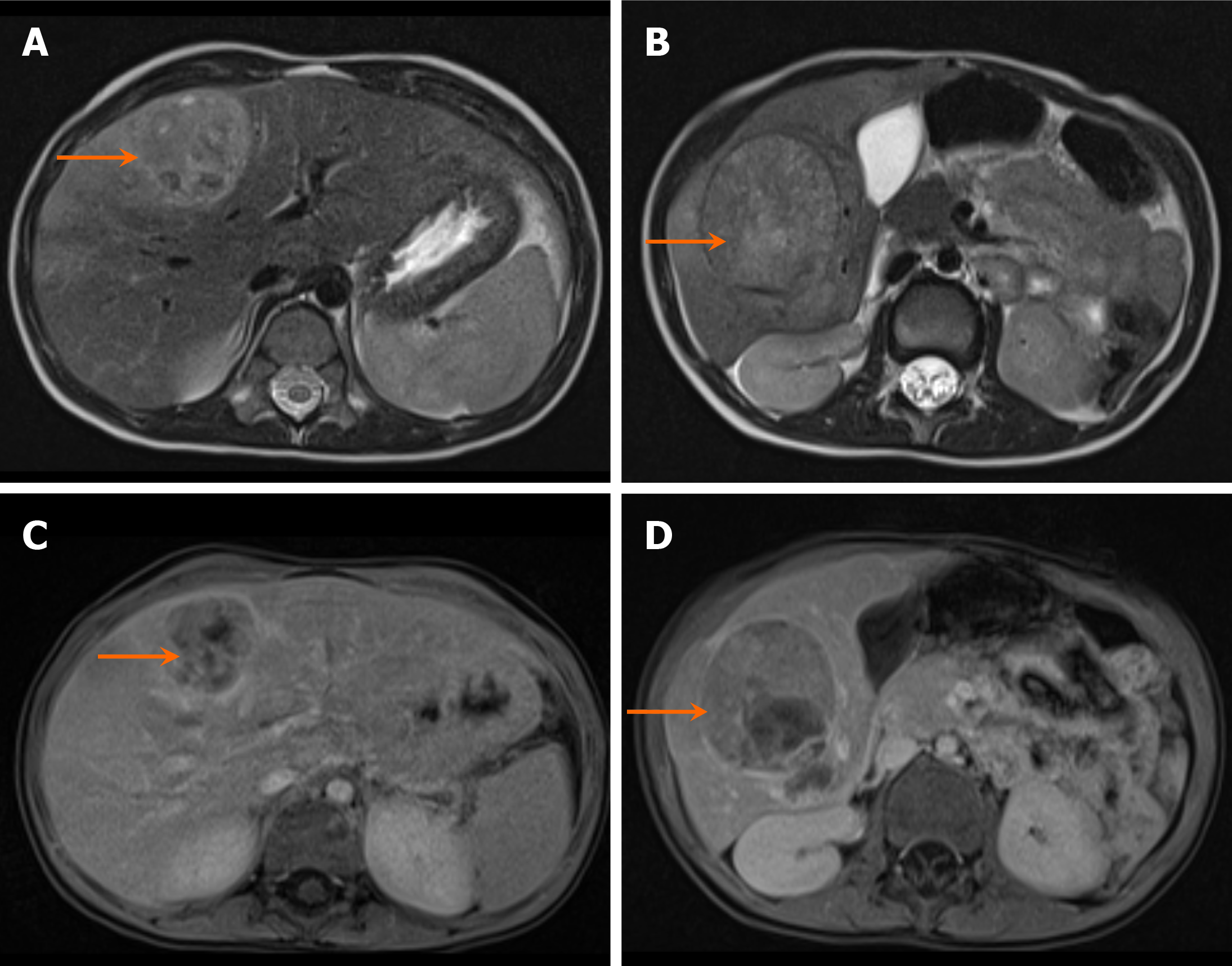
Figure 3 Hepatoblastoma in a two-year-old boy with an abdominal mass and increased alpha-fetoprotein levels.
A and B: Axial T2-weighted images from an magnetic resonance imaging abdomen show heterogeneous lesions (arrows) involving the right lobe of the liver, predominantly segments VI and V; C and D: Axial T1 post-contrast images. The lesions show heterogeneous postcontrast enhancement (arrows). There is no infiltration of the mass into the inferior vena cava, or portal or hepatic veins. This was staged as pretreatment extent of disease II F (multifocal) disease involving right anterior and posterior sections.
- Citation: Shahid M, Hilal K, Khan M, Ejaz ZH, Altaf S, Islam S, Khandwala K. Imaging insights into pediatric liver masses: A comprehensive minireview for hepatology practice. World J Hepatol 2025; 17(8): 107041
- URL: https://www.wjgnet.com/1948-5182/full/v17/i8/107041.htm
- DOI: https://dx.doi.org/10.4254/wjh.v17.i8.107041


