
















©The Author(s) 2025.
World J Hepatol. Aug 27, 2025; 17(8): 107041
Published online Aug 27, 2025. doi: 10.4254/wjh.v17.i8.107041
Published online Aug 27, 2025. doi: 10.4254/wjh.v17.i8.107041
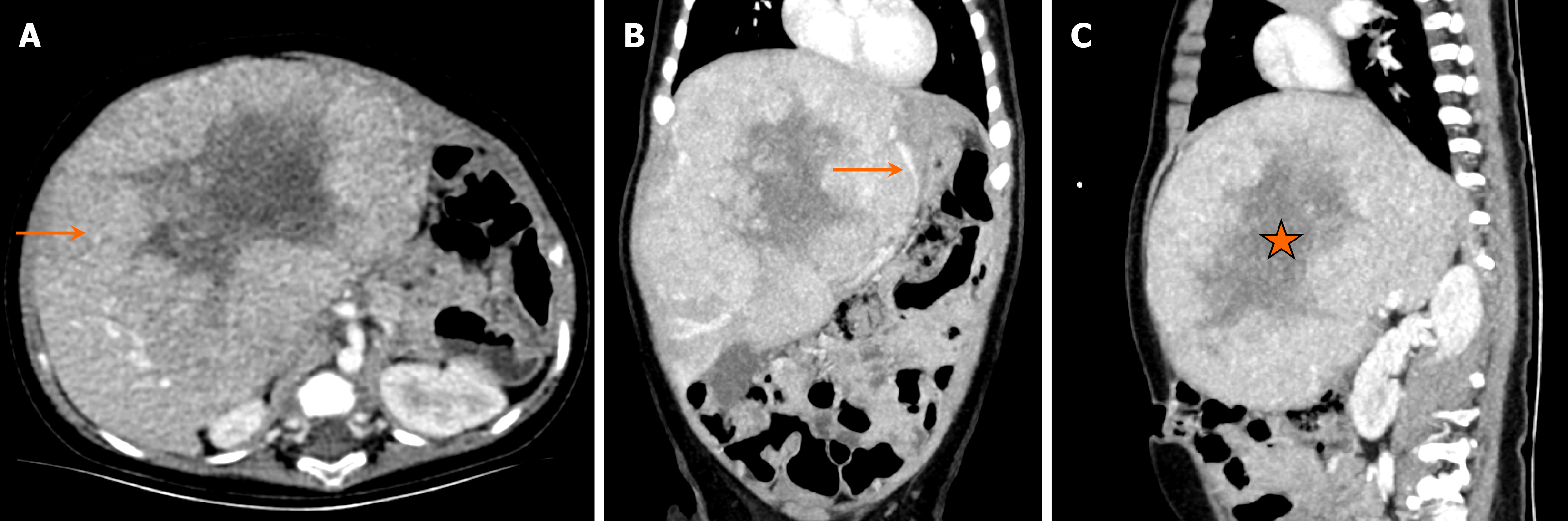
Figure 2 Hepatoblastoma in a six-month-old boy who presented with complaints of abdominal distension for two weeks.
A: Axial computed tomography (CT) image; B: Coronal CT image; C: Sagittal CT image. They show an enlarged liver with a large heterogeneous lesion occupying both lobes of the liver (arrow in A), involving segments I, IV, V and VIII. It displaces the left portal vein (arrow in B) and has internal low attenuation areas representing necrosis (asterisk in image C). No internal calcifications are noted. This was staged as pretreatment extent of disease III C (caudate).
- Citation: Shahid M, Hilal K, Khan M, Ejaz ZH, Altaf S, Islam S, Khandwala K. Imaging insights into pediatric liver masses: A comprehensive minireview for hepatology practice. World J Hepatol 2025; 17(8): 107041
- URL: https://www.wjgnet.com/1948-5182/full/v17/i8/107041.htm
- DOI: https://dx.doi.org/10.4254/wjh.v17.i8.107041