Copyright
©The Author(s) 2025.
World J Hepatol. Jul 27, 2025; 17(7): 107378
Published online Jul 27, 2025. doi: 10.4254/wjh.v17.i7.107378
Published online Jul 27, 2025. doi: 10.4254/wjh.v17.i7.107378
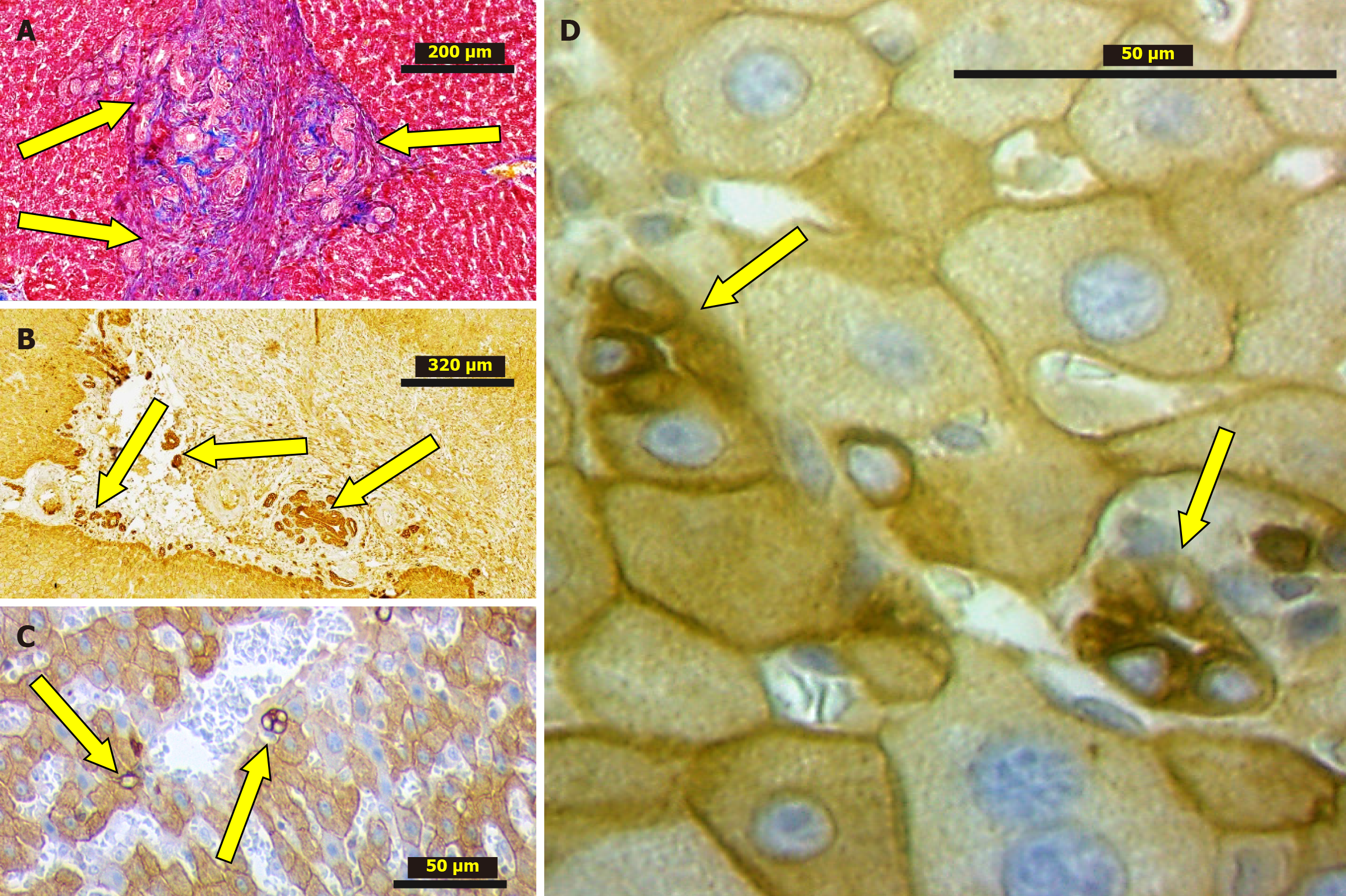
Figure 4 Ductular reaction (arrows).
A: The granulation tissue developed at the area of liver lobes adhesion after 2 weeks from partial hepatectomy (PH). Masson’s Trichrome; B: The granulation tissue developed at the area cut edge necrosis 2 weeks from PH. IHC stain, CK8; C and D: Hepatic lobules after 6 months from PH. IHC stain, CK8. Male Wistar rats.
- Citation: Korchilava B, Khachidze T, Megrelishvili N, Svanadze L, Kakabadze M, Tsomaia K, Jintcharadze M, Kordzaia D. Liver regeneration after partial hepatectomy: Triggers and mechanisms. World J Hepatol 2025; 17(7): 107378
- URL: https://www.wjgnet.com/1948-5182/full/v17/i7/107378.htm
- DOI: https://dx.doi.org/10.4254/wjh.v17.i7.107378


