Copyright
©The Author(s) 2025.
World J Hepatol. Jul 27, 2025; 17(7): 107378
Published online Jul 27, 2025. doi: 10.4254/wjh.v17.i7.107378
Published online Jul 27, 2025. doi: 10.4254/wjh.v17.i7.107378
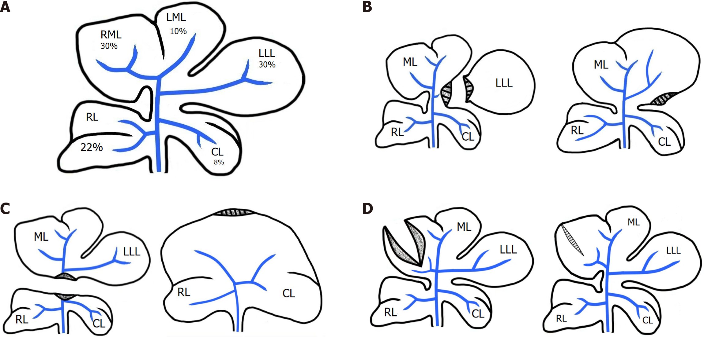
Figure 1 Schematic representation of rat liver lobes resection variations and Subsequent processes in the liver.
A: Normal rat liver; B: Resection of 30% of the liver mass – the Left Lateral Lobe (LLL) – followed by hypertrophic regeneration; C: Resection of 70% of the liver mass – the Right Median Lobe (RML), Left Median Lobe (LML), and LLL – followed by hyperplastic regeneration; D: Dissection of the median lobe (ML) and the subsequent healing and scar formation; RL: Right lateral lobe; CL: Caudate lobe; ML: Median lobe; LLL: Left lateral lobe; RML: Right Median Lobe, LML: Left Median Lobe.
- Citation: Korchilava B, Khachidze T, Megrelishvili N, Svanadze L, Kakabadze M, Tsomaia K, Jintcharadze M, Kordzaia D. Liver regeneration after partial hepatectomy: Triggers and mechanisms. World J Hepatol 2025; 17(7): 107378
- URL: https://www.wjgnet.com/1948-5182/full/v17/i7/107378.htm
- DOI: https://dx.doi.org/10.4254/wjh.v17.i7.107378


