Copyright
©The Author(s) 2025.
World J Hepatol. May 27, 2025; 17(5): 104646
Published online May 27, 2025. doi: 10.4254/wjh.v17.i5.104646
Published online May 27, 2025. doi: 10.4254/wjh.v17.i5.104646
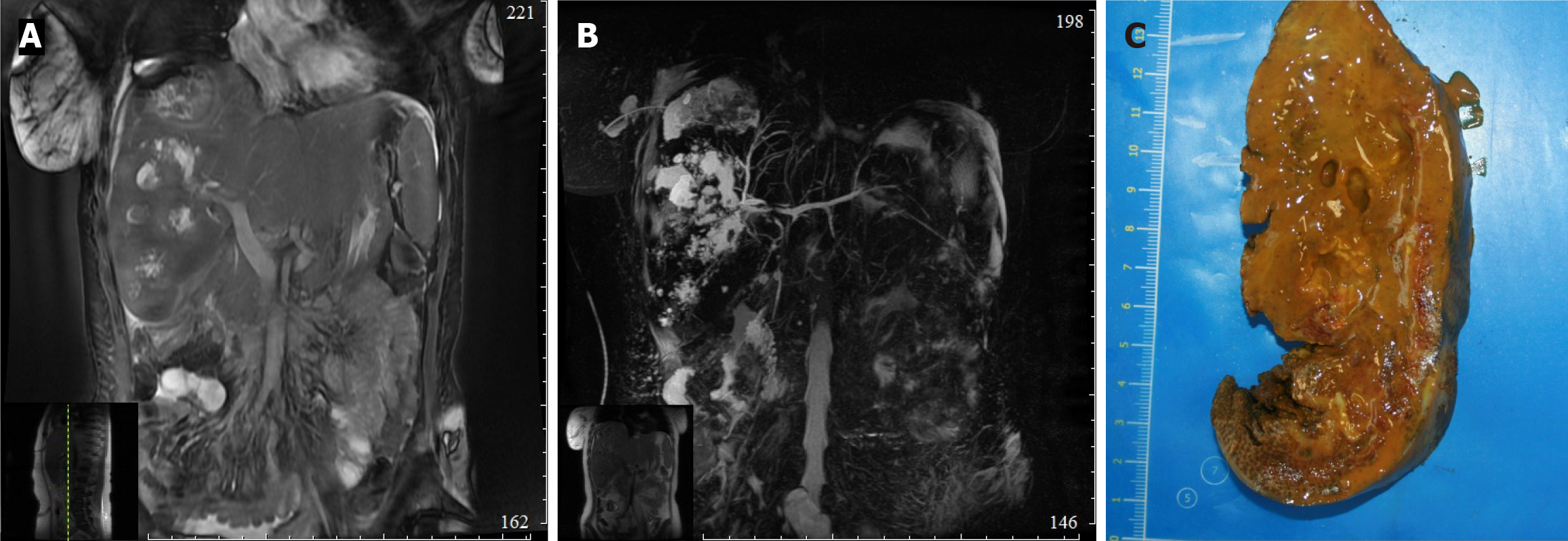
Figure 2 A clinical case of bile duct stricture.
A: Magnetic resonance imaging of the liver: Radiological picture of multiple abscesses of the right lobe of the liver, occlusion of the right hepatic vein; B: Magnetic resonance imaging of the liver and bile ducts: Radiological picture of multiple abscesses of the right lobe of the liver, stricture of the common hepatic duct; C: Macrophotograph: Merging areas of necrosis in the liver tissue with formation of large cavities with necrotic debris and bile in the lumen.
- Citation: Trifonov S, Kovalenko Y, Gurmikov B, Varava A, Vodeiko V, Pakhtushkin E, Vishnevsky V, Zharikov Y. Reconstructive surgery and percutaneous balloon dilation for the treatment of benign biliary strictures: A retrospective study. World J Hepatol 2025; 17(5): 104646
- URL: https://www.wjgnet.com/1948-5182/full/v17/i5/104646.htm
- DOI: https://dx.doi.org/10.4254/wjh.v17.i5.104646


